Improper dihedrals are a measure of the chirality/planarity of the structure at a specific atom. Values around -35 or +35 are expected for chiral atoms, and values around 0 for planar atoms. Planar side chains are left out of the calculations, these are better handled by the planarity checks.
Three numbers are given for each atom in the table. The first is the Z-score for the improper dihedral. The second number is the measured improper dihedral. The third number is the expected value for this atom type. A final column contains an extra warning if the chirality for an atom is opposite to the expected value.
429 ILE ( 429 ) C -4.4 -8.9 -0.2
Improper dihedral RMS Z-score : 0.556
Note: Chain names are OK
All chain names assigned to polymer molecules are unique, and all
residue numbers are strictly increasing within each chain.
Note: Weights checked OK
All atomic occupancy factors ('weights') fall in the 0.0--1.0 range.
Geometric checks
Note: No missing atoms detected
All expected atoms are present.
Note: OXT check OK
All required C-terminal oxygen atoms are present.
Note: No extra C-terminal groups found
No C-terminal groups are present for non C-terminal residues
Note: All bond lengths OK
All bond lengths are in agreement with standard bond lengths using
a tolerance of 4 sigma (both standard values and sigma for amino
acid residues have been taken from Engh and Huber [REF], for
DNA/RNA from Parkinson et al [REF])
Note: Normal bond length variability
Bond lengths were found to deviate normally from the standard bond
lengths (values for Protein residues were taken from Engh and Huber
[REF], for DNA/RNA from Parkinson et al [REF]).
RMS Z-score for bond lengths: 0.743
RMS-deviation in bond distances: 0.017
Note: No bond length directionality
Comparison of bond distances with Engh and Huber [REF] standard
values for protein residues and Parkinson et al [REF] values for
DNA/RNA does not show significant systematic deviations.
Warning: Unusual bond angles
The bond angles listed in the table below were found to deviate
more than 4 sigma from standard bond angles (both standard values
and sigma for protein residues have been taken from Engh and Huber
[REF], for DNA/RNA from Parkinson et al [REF]). In the table below
for each strange angle the bond angle and the number of standard
deviations it differs from the standard values is given. Please
note that disulphide bridges are neglected. Atoms starting with "<"
belong to the previous residue in the sequence.
213 PHE ( 213 ) CA CB CG 109.761 -4.0
RMS Z-score for bond angles: 0.904
RMS-deviation in bond angles: 1.813
Note: Side chain planarity OK
All of the side chains of residues that have a planar group are
planar within expected RMS deviations.
Note: Atoms connected to aromatic rings OK
All of the atoms that are connected to planar aromatic rings in side
chains of amino-acid residues are in the plane within expected RMS
deviations.
Warning: Unusual PRO puckering amplitudes
The proline residues listed in the table below have a puckering
amplitude that is outside of normal ranges. Puckering parameters
were calculated by the method of Cremer and Pople [REF]. Normal PRO
rings have a puckering amplitude Q between 0.20 and 0.45
Angstrom. If Q is lower than 0.20 Angstrom for a PRO residue, this
could indicate disorder between the two different normal ring forms
(with C-gamma below and above the ring, respectively). If Q is
higher than 0.45 Angstrom something could have gone wrong during the
refinement.
   4 PRO  (   4 )     0.19 LOW
 107 PRO  ( 107 )     0.12 LOW
 196 PRO  ( 196 )     0.10 LOW
 236 PRO  ( 236 )     0.16 LOW
 265 PRO  ( 265 )     0.15 LOW
  11 PRO  (  11 )    105.8 envelop C-beta (108 degrees)
 348 PRO  ( 348 )    104.6 envelop C-beta (108 degrees)
 416 PRO  ( 416 )     99.8 envelop C-beta (108 degrees)
These scores give an impression of how ``normal'' the torsion angles in protein residues are. All torsion angles except omega are used for calculating a `normality' score. Average values and standard deviations were obtained from the residues in the WHAT IF database. These are used to calculate Z-scores. A residue with a Z-score of below -2.0 is poor, and a score of less than -3.0 is worrying. For such residues more than one torsion angle is in a highly unlikely position.
 363 TRP  ( 363 )   -3.2262
 348 PRO  ( 348 )   -2.8409
 352 THR  ( 352 )   -2.7929
 147 SER  ( 147 )   -2.5771
 237 THR  ( 237 )   -2.4351
 393 LEU  ( 393 )   -2.3732
  10 GLU  (  10 )   -2.3492
 199 VAL  ( 199 )   -2.2300
 276 ARG  ( 276 )   -2.2219
 228 ASP  ( 228 )   -2.2054
 211 PRO  ( 211 )   -2.1579
 385 VAL  ( 385 )   -2.1332
  48 GLN  (  48 )   -2.1298
 326 ILE  ( 326 )   -2.1244
 392 GLY  ( 392 )   -2.1033
 225 GLU  ( 225 )   -2.0838
 426 LEU  ( 426 )   -2.0738
 183 LEU  ( 183 )   -2.0512
Residues with ``forbidden'' phi-psi combinations are listed, as well as residues with unusual omega angles (deviating by more than 3 sigma from the normal value). Please note that it is normal if about 5 percent of the residues is listed here as having unusual phi-psi combinations.
  10 GLU  (  10 )   Poor phi/psi
 158 THR  ( 158 )   Poor phi/psi
 161 ARG  ( 161 )   Poor phi/psi
 210 LEU  ( 210 )   PRO omega poor
 211 PRO  ( 211 )   Poor PRO-phi
 226 GLU  ( 226 )   Poor phi/psi
 228 ASP  ( 228 )   Poor phi/psi
 229 THR  ( 229 )   Poor phi/psi
 256 LEU  ( 256 )   PRO omega poor
 257 PRO  ( 257 )   Poor PRO-phi
 275 GLY  ( 275 )   Poor phi/psi
 276 ARG  ( 276 )   Poor phi/psi
 279 ARG  ( 279 )   Poor phi/psi
 280 GLY  ( 280 )   Poor phi/psi
 348 PRO  ( 348 )   Poor PRO-phi
 349 ALA  ( 349 )   Poor phi/psi
 356 ILE  ( 356 )   Poor phi/psi
 363 TRP  ( 363 )   Poor phi/psi
 371 GLN  ( 371 )   Poor phi/psi
 377 LYS  ( 377 )   Poor phi/psi
 380 LYS  ( 380 )   Poor phi/psi
 392 GLY  ( 392 )   omega poor
 425 GLY  ( 425 )   Poor phi/psi
Ramachandran Z-score : -1.754
Warning: Omega angles too tightly restrained
The omega angles for trans-peptide bonds in a structure are
expected to give a gaussian distribution with the average around
+178 degrees and a standard deviation around 5.5 degrees. These
expected values were obtained from very accurately determined
structures. Many protein structures are too tightly constrained.
This seems to be the case with the current structure, as the
observed standard deviation is below 4.0 degrees.
Standard deviation of omega values : 3.489
Note: chi-1/chi-2 angle correlation Z-score OK
The score expressing how well the chi-1/chi-2 angles of all residues
are corresponding to the populated areas in the database is
within expected ranges for well-refined structures.
chi-1/chi-2 correlation Z-score : -2.398
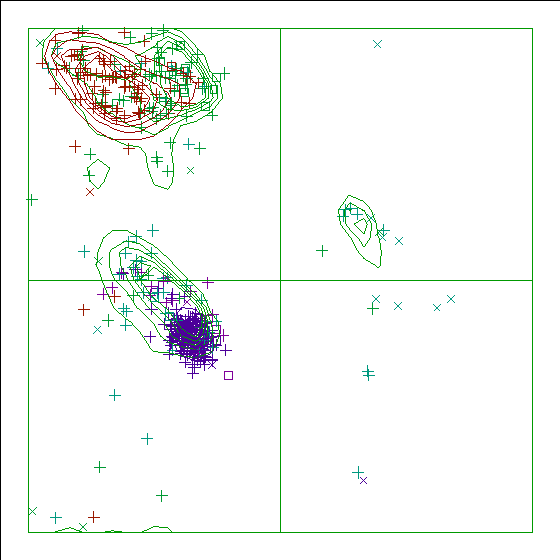
Note: Ramachandran plot
In this Ramachandran plot X-signs represent glycines, squares represent
prolines and small plus-signs represent the other residues. If too many
plus-signs fall outside the contoured areas then the molecule is poorly
refined (or worse).
In a colour picture, the residues that are part of a helix are shown in blue, strand residues in red. "Allowed" regions for helical residues are drawn in blue, for strand residues in red, and for all other residues in green.
Chain without chain identifier
Accessibility related checks
Note: Inside/Outside residue distribution normal
The distribution of residue types over the inside and the outside of the
protein is normal.
inside/outside RMS Z-score : 1.009
Note: Inside/Outside RMS Z-score plot
The Inside/Outside distribution normality RMS Z-score over a 15
residue window is plotted as function of the residue number. High
areas in the plot (above 1.5) indicate unusual inside/outside
patterns.
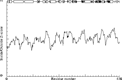
Chain without chain identifier
Secondary structure
Note: Secondary structure
This is the secondary structure according to DSSP. Only helix (H), strand
(S), turn (T) and coil (blank) are shown. [REF]
DBG> SSBOND cards to be written: 0
                     10        20        30        40        50        60
                      |         |         |         |         |         |
    1 -  60  MLDPNLLRNEPDAVAEKLARRGFKLDVDKLGALEERRKVLQVKTENLQAERNSRSKSIGQ
    1 -  60     HHHHHH HHHHHHHHHTTT    HHHHHHHHHHHHHHHHHHHHHHHHHHHHHHHHHH
                     70        80        90       100       110       120
                      |         |         |         |         |         |
   61 - 120  AKARGEDIEPLRLEVNKLGEELDAAKAELDALQAEIRDIALTIPNLPADEVPVGKDENDN
   61 - 120  HHHTT   HHHHHHHHHHHHHHHHHHHHHHHHHHHHHHHHHT      TTT   TT333
                    130       140       150       160       170       180
                      |         |         |         |         |         |
  121 - 180  VEVSRWGTPREFDFEVRDHVTLGEMHSGLDFAAAVKLTGSRFVVMKGQIARMHRALSQFM
  121 - 180  SSSSSST     TT    HHHHHHHTT SSHHHHHHHT TT  SS HHHHHHHHHHHHHH
                    190       200       210       220       230       240
                      |         |         |         |         |         |
  181 - 240  LDLHTEQHGYSENYVPYLVNQDTLYGTGQLPKFAGDLFHTRPLEEEADTSNYALIPTAEV
  181 - 240  HHHHHHTT  SS    TSSSHHHHHHHT TTTT333T SS    TT     SSS TTTHH
                    250       260       270       280       290       300
                      |         |         |         |         |         |
  241 - 300  PLTNLVRGEIIDEDDLPIKMTAHTPCFRSEAGSYGRDTRGLIRMHQFDKVEMVQIVRPED
  241 - 300  HHHHTTTT SSS333  SSSSSSSSSS T      TT  T    TSSSSSSSSSSS 333
                    310       320       330       340       350       360
                      |         |         |         |         |         |
  301 - 360  SMAALEEMTGHAEKVLQLLGLPYRKIILCTGDMGFGACKTYDLEVWIPAQNTYREISSCS
  301 - 360  HHHHHHHHHHHHHHHHHHHT  SSSSS  333T TT TSSSSSSSSS333TSSSSSSSSS
                    370       380       390       400       410       420
                      |         |         |         |         |         |
  361 - 420  NVWDFQARRMQARCRSKSDKKTRLVHTLNGSGLAVGRTLVAVMENYQQADGRIEVPEVLR
  361 - 420  S TTHHHHHHT SSS TTT  SSS SSSSSSSSSHHHHHHHHHHHT  TTT SS  TTT3
                    430
                      |
  421 - 430  PYMNGLEYIG
  421 - 430  33TTT  SS
The contact distances of all atom pairs have been checked. Two atoms are said to `bump' if they are closer than the sum of their Van der Waals radii minus 0.40 Angstrom. For hydrogen bonded pairs a tolerance of 0.55 Angstrom is used. The first number in the table tells you how much shorter that specific contact is than the acceptable limit. The second distance is the distance between the centers of the two atoms.
The last text-item on each line represents the status of the atom pair. The text `INTRA' means that the bump is between atoms that are explicitly listed in the PDB file. `INTER' means it is an inter-symmetry bump. If the final column contains the text 'HB', the bump criterium was relaxed because there could be a hydrogen bond. Similarly relaxed criteria are used for 1--3 and 1--4 interactions (listed as 'B2' and 'B3', respectively). If the last column is 'BF', the sum of the B-factors of the atoms is higher than 80, which makes the appearance of the bump somewhat less severe because the atoms probably aren't there anyway.
Bumps between atoms for which the sum of their occupancies is lower than one are not reported. In any case, each bump is listed in only one direction.
 305 LEU  ( 305 )      O    --  309 THR  ( 309 )      CG2    0.341   2.459 INTRA
 149 LEU  ( 149 )      CD2  --  151 PHE  ( 151 )      CE2    0.280   2.920 INTRA
 308 MET  ( 308 )      CE   --  388 LEU  ( 388 )      CB     0.274   2.926 INTRA
  10 GLU  (  10 )      N    --   11 PRO  (  11 )      CD     0.261   2.739 INTRA BF
 425 GLY  ( 425 )      O    --  426 LEU  ( 426 )      C      0.260   2.540 INTRA
 203 THR  ( 203 )      CG2  --  244 ASN  ( 244 )      ND2    0.239   2.861 INTRA
 387 THR  ( 387 )      C    --  388 LEU  ( 388 )      CD2    0.235   2.965 INTRA
  10 GLU  (  10 )      O    --   11 PRO  (  11 )      C      0.232   2.568 INTRA BF
 329 CYS  ( 329 )      O    --  330 THR  ( 330 )      C      0.229   2.571 INTRA
 415 VAL  ( 415 )      O    --  416 PRO  ( 416 )      C      0.215   2.585 INTRA
 326 ILE  ( 326 )      CD1  --  328 LEU  ( 328 )      CD2    0.209   2.991 INTRA
 161 ARG  ( 161 )      CZ   --  282 ILE  ( 282 )      CD1    0.207   2.993 INTRA BF
 263 HIS  ( 263 )      C    --  264 THR  ( 264 )      CG2    0.180   3.020 INTRA
 398 THR  ( 398 )      O    --  402 VAL  ( 402 )      CG1    0.178   2.622 INTRA
 348 PRO  ( 348 )      O    --  350 GLN  ( 350 )      N      0.165   2.535 INTRA
 361 ASN  ( 361 )      ND2  --  363 TRP  ( 363 )      N      0.162   2.838 INTRA
 420 ARG  ( 420 )      NH1  --  427 GLU  ( 427 )      CG     0.157   2.943 INTRA BF
  69 GLU  (  69 )      N    --   70 PRO  (  70 )      CD     0.156   2.844 INTRA BF
 192 GLU  ( 192 )      C    --  193 ASN  ( 193 )      ND2    0.154   2.946 INTRA
 184 HIS  ( 184 )      ND1  --  188 HIS  ( 188 )      CD2    0.144   2.956 INTRA
 128 THR  ( 128 )      O    --  129 PRO  ( 129 )      C      0.132   2.668 INTRA
 366 GLN  ( 366 )      O    --  367 ALA  ( 367 )      C      0.131   2.669 INTRA
 207 THR  ( 207 )      OG1  --  209 GLN  ( 209 )      NE2    0.124   2.426 INTRA HB
 171 ARG  ( 171 )      NH1  --  422 TYR  ( 422 )      O      0.123   2.577 INTRA
 246 VAL  ( 246 )      O    --  247 ARG  ( 247 )      C      0.112   2.688 INTRA
And so on for a total of 81 lines
The packing environment of the residues is compared with the average packing environment for all residues of the same type in good PDB files. A low packing score can indicate one of several things: Poor packing, misthreading of the sequence through the density, crystal contacts, contacts with a co-factor, or the residue is part of the active site. It is not uncommon to see a few of these, but in any case this requires further inspection of the residue.
 194 TYR  ( 194 )    -8.55
 279 ARG  ( 279 )    -7.22
 223 LEU  ( 223 )    -6.65
 283 ARG  ( 283 )    -6.13
 350 GLN  ( 350 )    -5.57
   9 ASN  (   9 )    -5.38
 115 LYS  ( 115 )    -5.37
 276 ARG  ( 276 )    -5.18
 125 ARG  ( 125 )    -5.15
 225 GLU  ( 225 )    -5.12
The table below lists the first and last residue in each stretch found, as well as the average residue score of the series.
223 LEU ( 223 ) --- 225 GLU ( 225 ) -5.28
Average for range 1 - 430 : -0.760
Note: Quality value plot
The quality value smoothed over a 10 residue window is plotted as
function of the residue number. Low areas in the plot (below
-2.0) indicate "unusual" packing.
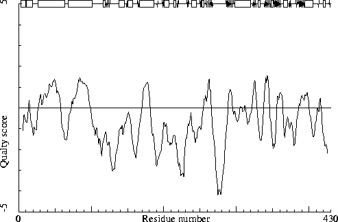
Chain without chain identifier
Warning: Low packing Z-score for some residues
The residues listed in the table below have an unusual packing
environment according to the 2nd generation quality check. The score
listed in the table is a packing normality Z-score: positive means
better than average, negative means worse than average. Only residues
scoring less than -2.50 are listed here. These are the "unusual"
residues in the structure, so it will be interesting to take a
special look at them.
 134 PHE  ( 134 )    -2.94
 194 TYR  ( 194 )    -2.59
 222 PRO  ( 222 )    -2.58
The table below lists the first and last residue in each stretch found, as well as the average residue Z-score of the series.
279 ARG ( 279 ) --- 282 ILE ( 282 ) -2.10
All contacts : Average = -0.095 Z-score = -0.47
BB-BB contacts : Average = 0.164 Z-score = 1.19
BB-SC contacts : Average = -0.335 Z-score = -1.75
SC-BB contacts : Average = 0.009 Z-score = 0.23
SC-SC contacts : Average = -0.264 Z-score = -1.14
Note: Second generation quality Z-score plot
The second generation quality Z-score smoothed over a 10 residue window
is plotted as function of the residue number. Low areas in the plot (below
-1.3) indicate "unusual" packing.
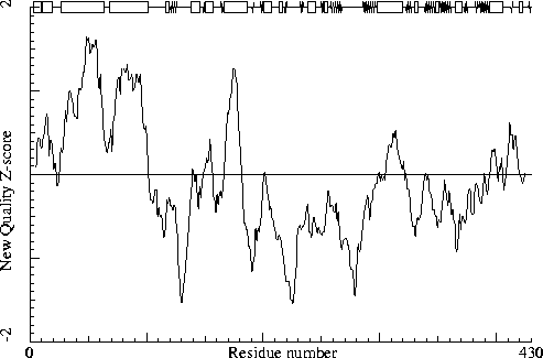
Chain without chain identifier
Note: Backbone oxygen evaluation OK
All residues for which the local backbone conformation could be
found in the WHAT IF database have a normal backbone oxygen
position.
Warning: Unusual rotamers
The residues listed in the table below have a rotamer that is not
seen very often in the database of solved protein structures. This
option determines for every residue the position specific chi-1
rotamer distribution. Thereafter it verified whether the actual
residue in the molecule has the most preferred rotamer or not. If
the actual rotamer is the preferred one, the score is 1.0. If the
actual rotamer is unique, the score is 0.0. If there are two
preferred rotamers, with a population distribution of 3:2 and your
rotamer sits in the lesser populated rotamer, the score will be
0.66. No value will be given if insufficient hits are found in the
database.
It is not necessarily an error if a few residues have rotamer values below 0.3, but careful inspection of all residues with these low values could be worth it.
 417 GLU  ( 417 )     0.33
 389 ASN  ( 389 )     0.38
 125 ARG  ( 125 )     0.38
For this check, backbone conformations are compared with database structures using C-alpha superpositions with some restraints on the backbone oxygen positions.
A residue mentioned in the table can be part of a strange loop, or there might be something wrong with it or its directly surrounding residues. There are a few of these in every protein, but in any case it is worth looking at!
 158 THR  ( 158 )    0
 210 LEU  ( 210 )    0
 228 ASP  ( 228 )    0
 229 THR  ( 229 )    0
 276 ARG  ( 276 )    0
 363 TRP  ( 363 )    0
 377 LYS  ( 377 )    0
 379 ASP  ( 379 )    0
 393 LEU  ( 393 )    0
 147 SER  ( 147 )    1
 166 LYS  ( 166 )    1
 226 GLU  ( 226 )    1
 256 LEU  ( 256 )    1
 364 ASP  ( 364 )    1
 134 PHE  ( 134 )    2
 161 ARG  ( 161 )    2
 168 GLN  ( 168 )    2
 224 GLU  ( 224 )    2
 227 ALA  ( 227 )    2
Backbone conformation Z-score : -0.153
B-factor analysis
Warning: Average B-factor out of normal range
The average B-factor for all buried protein atoms normally lies between
10--20. Values around 3--5 are expected for X-ray studies performed
at liquid nitrogen temperature.
Average B-factor for buried atoms : 25.038
Note: Number of buried atoms with low B-factor is OK
For protein structures determined at room temperature, no more than
about 1 percent of the B factors of buried atoms is below 5.0.
Percentage of buried atoms with B less than 5 : 0.26
Note: B-factor distribution normal
The distribution of B-factors within residues is within expected
ranges. A value over 1.5 here would mean that the B-factors show
signs of over-refinement.
RMS Z-score : 0.634 over 2836 bonds
Average difference in B over a bond : 1.84
RMS difference in B over a bond : 2.41
Note: B-factor plot
The average atomic B-factor per residue is plotted as function of
the residue number.
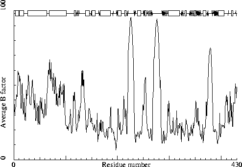
Chain without chain identifier
Hydrogen bond related checks
Error: HIS, ASN, GLN side chain flips
Listed here are Histidine, Asparagine or Glutamine residues for
which the orientation determined from hydrogen bonding analysis are
different from the assignment given in the input. Either they could
form energetically more favorable hydrogen bonds if the terminal
group was rotated by 180 degrees, or there is no assignment in the
input file (atom type 'A') but an assignment could be made. If a
residue is marked ``flexible'' the flipped conformation is only
slightly better than the non-flipped conformation.
 188 HIS  ( 188 )
 219 HIS  ( 219 )
 285 HIS  ( 285 )
 361 ASN  ( 361 )
In the table below all normal histidine residues are listed. The assignment based on the geometry of the residue is listed first, together with the RMS Z-score for the fit to the Engh and Huber parameters. For all residues where the H-bond assignment is different, the assignment is listed in the last columns, together with its RMS Z-score to the Engh and Huber parameters.
As always, the RMS Z-scores should be close to 1.0 if the residues were restrained to the Engh and Huber parameters during refinement.
Please note that because the differences between the geometries of the different types are small it is possible that the geometric assignment given here does not correspond to the type used in refinement. This is especially true if the RMS Z-scores are much higher than 1.0.
If the two assignments differ, or the ``geometry'' RMS Z-score is high, it is advisable to verify the hydrogen bond assignment, check the HIS type used during the refinement and possibly adjust it.
 139 HIS  ( 139 )     HIS-E   0.79 HIS-D   1.01
 146 HIS  ( 146 )     HIS-E   0.83
 173 HIS  ( 173 )     HIS-D   0.75 HIS-E   0.94
 184 HIS  ( 184 )     HIS-E   0.74
 188 HIS  ( 188 )     HIS-D   0.70
 219 HIS  ( 219 )     HIS-E   0.88
 263 HIS  ( 263 )     HIS-D   0.83 HIS-E   0.92
 285 HIS  ( 285 )     HIS-D   0.74
 311 HIS  ( 311 )     HIS-E   0.79
 386 HIS  ( 386 )     HIS-E   0.70
Hydrogen bond donors that are buried inside the protein normally use all of their hydrogens to form hydrogen bonds within the protein. If there are any non hydrogen bonded buried hydrogen bond donors in the structure they will be listed here. In very good structures the number of listed atoms will tend to zero.
   1 MET  (   1 )      N
  12 ASP  (  12 )      N
  20 ARG  (  20 )      NE
  37 ARG  (  37 )      NE
  44 THR  (  44 )      OG1
 120 ASN  ( 120 )      N
 136 VAL  ( 136 )      N
 140 VAL  ( 140 )      N
 162 PHE  ( 162 )      N
 212 LYS  ( 212 )      N
 218 PHE  ( 218 )      N
 226 GLU  ( 226 )      N
 237 THR  ( 237 )      N
 238 ALA  ( 238 )      N
 279 ARG  ( 279 )      NH2
 280 GLY  ( 280 )      N
 281 LEU  ( 281 )      N
 286 GLN  ( 286 )      N
 324 ARG  ( 324 )      NE
 340 THR  ( 340 )      N
 352 THR  ( 352 )      N
 353 TYR  ( 353 )      N
 364 ASP  ( 364 )      N
 366 GLN  ( 366 )      N
 367 ALA  ( 367 )      N
 369 ARG  ( 369 )      NE
 369 ARG  ( 369 )      NH2
 377 LYS  ( 377 )      N
 379 ASP  ( 379 )      N
 380 LYS  ( 380 )      N
 381 LYS  ( 381 )      N
 412 ARG  ( 412 )      NE
Side-chain hydrogen bond acceptors that are buried inside the protein normally form hydrogen bonds within the protein. If there are any not hydrogen bonded in the optimized hydrogen bond network they will be listed here.
291 GLU ( 291 ) OE1
The second part of the table mostly gives an impression of how well the model conforms to common refinement constraint values. The first part of the table shows a number of constraint-independent quality indicators.
Structure Z-scores, positive is better than average:
  1st generation packing quality :  -0.651
  2nd generation packing quality :  -0.473
  Ramachandran plot appearance   :  -1.754
  chi-1/chi-2 rotamer normality  :  -2.398
  Backbone conformation          :  -0.153
  Bond lengths                   :   0.743
  Bond angles                    :   0.904
  Omega angle restraints         :   0.634 (tight)
  Side chain planarity           :   0.063 (tight)
  Improper dihedral distribution :   0.556
  B-factor distribution          :   0.634
  Inside/Outside distribution    :   1.009