Improper dihedrals are a measure of the chirality/planarity of the structure at a specific atom. Values around -35 or +35 are expected for chiral atoms, and values around 0 for planar atoms. Planar side chains are left out of the calculations, these are better handled by the planarity checks.
Three numbers are given for each atom in the table. The first is the Z-score for the improper dihedral. The second number is the measured improper dihedral. The third number is the expected value for this atom type. A final column contains an extra warning if the chirality for an atom is opposite to the expected value.
424 ILE ( 429 ) C -4.4 -8.9 -0.2
Improper dihedral RMS Z-score : 0.528
Note: Chain names are OK
All chain names assigned to polymer molecules are unique, and all
residue numbers are strictly increasing within each chain.
Note: Weights checked OK
All atomic occupancy factors ('weights') fall in the 0.0--1.0 range.
Geometric checks
Note: No missing atoms detected
All expected atoms are present.
Note: OXT check OK
All required C-terminal oxygen atoms are present.
Note: No extra C-terminal groups found
No C-terminal groups are present for non C-terminal residues
Warning: Unusual bond lengths
The bond lengths listed in the table below were found to deviate
more than 4 sigma from standard bond lengths (both standard values
and sigma for amino acid residues have been taken from Engh and
Huber [REF], for DNA they were taken from Parkinson et al [REF]). In
the table below for each unusual bond the bond length and the
number of standard deviations it differs from the normal value is
given.
Atom names starting with "<" belong to the previous residue in the chain. If the second atom name is "--SS", the disulphide bridge has a deviating length.
 244 VAL  ( 249 )      N    CA    1.381  -4.0
 315 ASN  ( 320 )      N    CA    1.540   4.3
 316 LEU  ( 321 )      N    CA    1.374  -4.4
RMS Z-score for bond lengths: 0.766
RMS-deviation in bond distances: 0.017
Note: No bond length directionality
Comparison of bond distances with Engh and Huber [REF] standard
values for protein residues and Parkinson et al [REF] values for
DNA/RNA does not show significant systematic deviations.
Note: All bond angles OK
All bond angles are in agreement with standard bond angles using a
tolerance of 4 sigma (both standard values and sigma for protein
residues have been taken from Engh and Huber [REF], for DNA/RNA
from Parkinson et al. [REF]). Please note that only bond angles
within protein residues are taken into account: disulphide bridges
and peptide bonds are neglected.
Note: Normal bond angle variability
Bond angles were found to deviate normally from the mean standard
bond angles (normal values for protein residues were taken from
Engh and Huber [REF], for DNA/RNA from Parkinson et al [REF]). The
RMS Z-score given below is expected to be around 1.0 for a normally
restrained data set, and this is indeed observed for very high
resolution X-ray structures. More common values are around 1.55
RMS Z-score for bond angles: 0.886
RMS-deviation in bond angles: 1.783
Note: Side chain planarity OK
All of the side chains of residues that have a planar group are
planar within expected RMS deviations.
Note: Atoms connected to aromatic rings OK
All of the atoms that are connected to planar aromatic rings in side
chains of amino-acid residues are in the plane within expected RMS
deviations.
Warning: Unusual PRO puckering amplitudes
The proline residues listed in the table below have a puckering
amplitude that is outside of normal ranges. Puckering parameters
were calculated by the method of Cremer and Pople [REF]. Normal PRO
rings have a puckering amplitude Q between 0.20 and 0.45
Angstrom. If Q is lower than 0.20 Angstrom for a PRO residue, this
could indicate disorder between the two different normal ring forms
(with C-gamma below and above the ring, respectively). If Q is
higher than 0.45 Angstrom something could have gone wrong during the
refinement.
   4 PRO  (   4 )     0.17 LOW
 105 PRO  ( 107 )     0.13 LOW
 194 PRO  ( 196 )     0.13 LOW
 231 PRO  ( 236 )     0.16 LOW
 260 PRO  ( 265 )     0.15 LOW
 226 PRO  ( 228 )     -9.5 half-chair C-alpha/N (-18 degrees)
 343 PRO  ( 348 )    104.8 envelop C-beta (108 degrees)
 411 PRO  ( 416 )     99.8 envelop C-beta (108 degrees)
These scores give an impression of how ``normal'' the torsion angles in protein residues are. All torsion angles except omega are used for calculating a `normality' score. Average values and standard deviations were obtained from the residues in the WHAT IF database. These are used to calculate Z-scores. A residue with a Z-score of below -2.0 is poor, and a score of less than -3.0 is worrying. For such residues more than one torsion angle is in a highly unlikely position.
 358 TRP  ( 363 )   -3.2357
 343 PRO  ( 348 )   -2.8409
 347 THR  ( 352 )   -2.7967
 232 THR  ( 237 )   -2.4351
 191 THR  ( 193 )   -2.4106
 388 LEU  ( 393 )   -2.3841
 197 VAL  ( 199 )   -2.2844
 271 ARG  ( 276 )   -2.2219
 181 LEU  ( 183 )   -2.2050
 380 VAL  ( 385 )   -2.1332
  46 GLN  (  48 )   -2.1298
 387 GLY  ( 392 )   -2.1033
 122 LEU  ( 124 )   -2.0847
  22 MET  (  24 )   -2.0804
 421 LEU  ( 426 )   -2.0704
  42 THR  (  44 )   -2.0421
 419 GLY  ( 424 )   -2.0197
 209 PRO  ( 211 )   -2.0045
Residues with ``forbidden'' phi-psi combinations are listed, as well as residues with unusual omega angles (deviating by more than 3 sigma from the normal value). Please note that it is normal if about 5 percent of the residues is listed here as having unusual phi-psi combinations.
  20 ASN  (  22 )   Poor phi/psi
 156 ALA  ( 158 )   Poor phi/psi
 159 ARG  ( 161 )   Poor phi/psi
 208 LEU  ( 210 )   PRO omega poor
 209 PRO  ( 211 )   Poor PRO-phi
 224 GLU  ( 226 )   Poor phi/psi
 243 ASP  ( 248 )   Poor phi/psi
 251 LEU  ( 256 )   PRO omega poor
 252 PRO  ( 257 )   Poor PRO-phi
 270 GLY  ( 275 )   Poor phi/psi
 271 ARG  ( 276 )   Poor phi/psi
 274 ARG  ( 279 )   Poor phi/psi
 275 GLY  ( 280 )   Poor phi/psi
 343 PRO  ( 348 )   Poor PRO-phi
 344 ALA  ( 349 )   Poor phi/psi
 351 ILE  ( 356 )   Poor phi/psi
 358 TRP  ( 363 )   Poor phi/psi
 366 GLN  ( 371 )   Poor phi/psi
 372 LYS  ( 377 )   Poor phi/psi
 375 LYS  ( 380 )   Poor phi/psi
 387 GLY  ( 392 )   omega poor
 420 GLY  ( 425 )   Poor phi/psi
Ramachandran Z-score : -1.642
Warning: Omega angles too tightly restrained
The omega angles for trans-peptide bonds in a structure are
expected to give a gaussian distribution with the average around
+178 degrees and a standard deviation around 5.5 degrees. These
expected values were obtained from very accurately determined
structures. Many protein structures are too tightly constrained.
This seems to be the case with the current structure, as the
observed standard deviation is below 4.0 degrees.
Standard deviation of omega values : 3.391
Note: chi-1/chi-2 angle correlation Z-score OK
The score expressing how well the chi-1/chi-2 angles of all residues
are corresponding to the populated areas in the database is
within expected ranges for well-refined structures.
chi-1/chi-2 correlation Z-score : -2.083
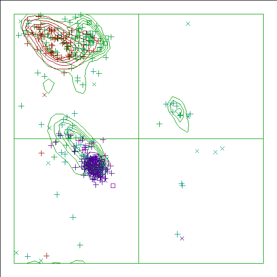
Note: Ramachandran plot
In this Ramachandran plot X-signs represent glycines, squares represent
prolines and small plus-signs represent the other residues. If too many
plus-signs fall outside the contoured areas then the molecule is poorly
refined (or worse).
In a colour picture, the residues that are part of a helix are shown in blue, strand residues in red. "Allowed" regions for helical residues are drawn in blue, for strand residues in red, and for all other residues in green.
Chain without chain identifier
Accessibility related checks
Note: Inside/Outside residue distribution normal
The distribution of residue types over the inside and the outside of the
protein is normal.
inside/outside RMS Z-score : 1.029
Note: Inside/Outside RMS Z-score plot
The Inside/Outside distribution normality RMS Z-score over a 15
residue window is plotted as function of the residue number. High
areas in the plot (above 1.5) indicate unusual inside/outside
patterns.
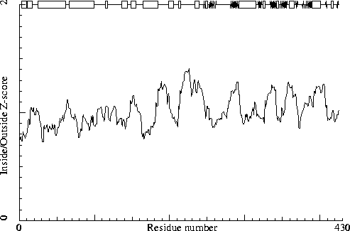
Chain without chain identifier
Secondary structure
Note: Secondary structure
This is the secondary structure according to DSSP. Only helix (H), strand
(S), turn (T) and coil (blank) are shown. [REF]
DBG> SSBOND cards to be written: 0
DBG> SSBOND cards to be written: 0
DBG> SSBOND cards to be written: 0
                     10
                      |
    1 -  18  MIDPNLLRNNLAEVAEKL
    1 -  18     HHHHHH HHHHHHH
             20        30        40        50        60        70
              |         |         |         |         |         |
   19 -  78  RNFMLDTEKLTALEDQRKNLQVTTENLQAERNARSKAIGAAKARGEDIAPLLAEMDDMGN
   19 -  78        HHHHHHHHHHHHHHHHHHHHHHHHHHHHHHHHHHHHHTT   HHHHHHHHHHHH
             80        90       100       110       120       130
              |         |         |         |         |         |
   79 - 138  QLTEAKAQLDAVLAEINQIALSIPNLPADEVPLGKDDTENKEILRWGTPRTFDFEVKDHI
   79 - 138  HHHHHHHHHHHHHHHHHHHHHT      TTT   TT333       T     TT    HH
            140       150       160       170       180       190
              |         |         |         |         |         |
  139 - 198  TLGEEANGLDFAAGAKLAGARFAVMKGQIAKMHRALAQFMLDLHTEQHGYLETYVPYLVN
  139 - 198  HHHHHTT   HHHHHHHT TT     HHHHHHHHHHHHHHHHHHHHTT        TT
            200       210       220
              |         |         |
  199 - 226  HATLYGTGQLPKFGEDLFHTLALEGEQP
  199 - 226  HHHHHHHT TTTT333T       TT
              230       240       250       260       270       280
                |         |         |         |         |         |
  227 - 286  YALIPTAEVPVTNLVRDVIIDEAELPIKMTAHTPCFRSEAGSYGRDTRGLIRMHQFDKVE
  227 - 286      TTTHHHHHHTTTT SSS333  SSSSSSSSSS T      TT  T    TSSSSSS
              290       300       310       320       330       340
                |         |         |         |         |         |
  287 - 346  MVQIVDPDKSMEALEELTGHAEKVLQLLNLPYRKVLLCTGDMGFGSCKTYDLEVWVPAQN
  287 - 346  SSSSS 333HHHHHHHHHHHHHHHHHHHT  SSSSS  333T TT TSSSSSSSSS333T
              350       360       370       380       390       400
                |         |         |         |         |         |
  347 - 406  TYREISSCSNMWDFQARRMQARCKAKGDKKTRLVHTLNGSGLAVGRTLVAVLENYQNADG
  347 - 406  SSSSSSSSSS TTHHHHHHT SSS TTT  SSS SSSSSSSSSHHHHHHHHHHHT  TTT
              410       420
                |         |
  407 - 425  SITVPEELRPYMGGLDVIG
  407 - 425   SS  TTT333TTT  SS
The contact distances of all atom pairs have been checked. Two atoms are said to `bump' if they are closer than the sum of their Van der Waals radii minus 0.40 Angstrom. For hydrogen bonded pairs a tolerance of 0.55 Angstrom is used. The first number in the table tells you how much shorter that specific contact is than the acceptable limit. The second distance is the distance between the centers of the two atoms.
The last text-item on each line represents the status of the atom pair. The text `INTRA' means that the bump is between atoms that are explicitly listed in the PDB file. `INTER' means it is an inter-symmetry bump. If the final column contains the text 'HB', the bump criterium was relaxed because there could be a hydrogen bond. Similarly relaxed criteria are used for 1--3 and 1--4 interactions (listed as 'B2' and 'B3', respectively). If the last column is 'BF', the sum of the B-factors of the atoms is higher than 80, which makes the appearance of the bump somewhat less severe because the atoms probably aren't there anyway.
Bumps between atoms for which the sum of their occupancies is lower than one are not reported. In any case, each bump is listed in only one direction.
 311 LEU  ( 316 )      O    --  315 ASN  ( 320 )      N      0.604   1.946 INTRA HB
 247 ASP  ( 252 )      O    --  250 GLU  ( 255 )      CG     0.339   2.461 INTRA
 300 LEU  ( 305 )      O    --  304 THR  ( 309 )      CG2    0.330   2.470 INTRA
  10 ASN  (  10 )      O    --   11 LEU  (  11 )      C      0.310   2.490 INTRA BF
 311 LEU  ( 316 )      O    --  315 ASN  ( 320 )      CA     0.301   2.499 INTRA
 420 GLY  ( 425 )      O    --  421 LEU  ( 426 )      C      0.265   2.535 INTRA
 312 GLN  ( 317 )      O    --  315 ASN  ( 320 )      N      0.253   2.447 INTRA
 237 VAL  ( 242 )      CG1  --  288 VAL  ( 293 )      CG2    0.246   2.954 INTRA
 147 LEU  ( 149 )      CD2  --  149 PHE  ( 151 )      CE2    0.238   2.962 INTRA
 201 THR  ( 203 )      CG2  --  239 ASN  ( 244 )      ND2    0.237   2.863 INTRA
 382 THR  ( 387 )      C    --  383 LEU  ( 388 )      CD2    0.234   2.966 INTRA
 324 CYS  ( 329 )      O    --  325 THR  ( 330 )      C      0.228   2.572 INTRA
 410 VAL  ( 415 )      O    --  411 PRO  ( 416 )      C      0.215   2.585 INTRA
 159 ARG  ( 161 )      CZ   --  277 ILE  ( 282 )      CD1    0.207   2.993 INTRA BF
 356 ASN  ( 361 )      ND2  --  358 TRP  ( 363 )      N      0.188   2.812 INTRA
 124 TRP  ( 126 )      CH2  --  315 ASN  ( 320 )      CB     0.187   3.013 INTRA
 393 THR  ( 398 )      O    --  397 VAL  ( 402 )      CG1    0.180   2.620 INTRA
 258 HIS  ( 263 )      C    --  259 THR  ( 264 )      CG2    0.180   3.020 INTRA
   9 ASN  (   9 )      O    --   10 ASN  (  10 )      ND2    0.176   2.524 INTRA
 343 PRO  ( 348 )      O    --  345 GLN  ( 350 )      N      0.167   2.533 INTRA
  67 ALA  (  69 )      N    --   68 PRO  (  70 )      CD     0.166   2.834 INTRA BF
  11 LEU  (  11 )      CB   --   25 THR  (  27 )      CG2    0.160   3.040 INTRA
   9 ASN  (   9 )      O    --   10 ASN  (  10 )      CG     0.158   2.642 INTRA
 182 HIS  ( 184 )      ND1  --  186 HIS  ( 188 )      CD2    0.157   2.943 INTRA
 322 LEU  ( 327 )      CD1  --  336 TYR  ( 341 )      CE1    0.156   3.044 INTRA
And so on for a total of 113 lines
The packing environment of the residues is compared with the average packing environment for all residues of the same type in good PDB files. A low packing score can indicate one of several things: Poor packing, misthreading of the sequence through the density, crystal contacts, contacts with a co-factor, or the residue is part of the active site. It is not uncommon to see a few of these, but in any case this requires further inspection of the residue.
 192 TYR  ( 194 )    -8.61
 225 GLN  ( 227 )    -7.24
 274 ARG  ( 279 )    -7.22
 221 LEU  ( 223 )    -6.79
 278 ARG  ( 283 )    -6.14
 111 LEU  ( 113 )    -5.77
 345 GLN  ( 350 )    -5.58
 113 LYS  ( 115 )    -5.40
 271 ARG  ( 276 )    -5.18
 123 ARG  ( 125 )    -5.08
The table below lists the first and last residue in each stretch found, as well as the average residue score of the series.
224 GLU ( 226 ) --- 226 PRO ( 228 ) -5.44
Average for range 1 - 425 : -0.753
Note: Quality value plot
The quality value smoothed over a 10 residue window is plotted as
function of the residue number. Low areas in the plot (below
-2.0) indicate "unusual" packing.
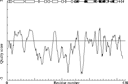
Chain without chain identifier
Warning: Low packing Z-score for some residues
The residues listed in the table below have an unusual packing
environment according to the 2nd generation quality check. The score
listed in the table is a packing normality Z-score: positive means
better than average, negative means worse than average. Only residues
scoring less than -2.50 are listed here. These are the "unusual"
residues in the structure, so it will be interesting to take a
special look at them.
 192 TYR  ( 194 )    -2.61
 186 HIS  ( 188 )    -2.52
The table below lists the first and last residue in each stretch found, as well as the average residue Z-score of the series.
274 ARG ( 279 ) --- 277 ILE ( 282 ) -2.12
All contacts : Average = -0.069 Z-score = -0.30
BB-BB contacts : Average = 0.191 Z-score = 1.38
BB-SC contacts : Average = -0.369 Z-score = -1.94
SC-BB contacts : Average = 0.095 Z-score = 0.75
SC-SC contacts : Average = -0.222 Z-score = -0.90
Note: Second generation quality Z-score plot
The second generation quality Z-score smoothed over a 10 residue window
is plotted as function of the residue number. Low areas in the plot (below
-1.3) indicate "unusual" packing.
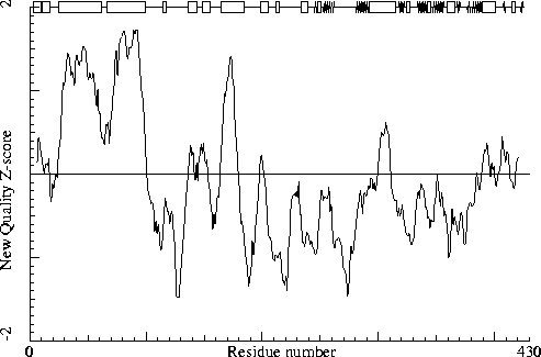
Chain without chain identifier
Note: Backbone oxygen evaluation OK
All residues for which the local backbone conformation could be
found in the WHAT IF database have a normal backbone oxygen
position.
Warning: Unusual rotamers
The residues listed in the table below have a rotamer that is not
seen very often in the database of solved protein structures. This
option determines for every residue the position specific chi-1
rotamer distribution. Thereafter it verified whether the actual
residue in the molecule has the most preferred rotamer or not. If
the actual rotamer is the preferred one, the score is 1.0. If the
actual rotamer is unique, the score is 0.0. If there are two
preferred rotamers, with a population distribution of 3:2 and your
rotamer sits in the lesser populated rotamer, the score will be
0.66. No value will be given if insufficient hits are found in the
database.
It is not necessarily an error if a few residues have rotamer values below 0.3, but careful inspection of all residues with these low values could be worth it.
 412 GLU  ( 417 )     0.33
 384 ASN  ( 389 )     0.38
For this check, backbone conformations are compared with database structures using C-alpha superpositions with some restraints on the backbone oxygen positions.
A residue mentioned in the table can be part of a strange loop, or there might be something wrong with it or its directly surrounding residues. There are a few of these in every protein, but in any case it is worth looking at!
 156 ALA  ( 158 )    0
 208 LEU  ( 210 )    0
 271 ARG  ( 276 )    0
 314 LEU  ( 319 )    0
 358 TRP  ( 363 )    0
 372 LYS  ( 377 )    0
 374 ASP  ( 379 )    0
 388 LEU  ( 393 )    0
 132 PHE  ( 134 )    1
 164 LYS  ( 166 )    1
 224 GLU  ( 226 )    1
 251 LEU  ( 256 )    1
 166 GLN  ( 168 )    2
 222 GLU  ( 224 )    2
 357 MET  ( 362 )    2
Backbone conformation Z-score : -0.169
B-factor analysis
Note: Average B-factor OK
The average B-factor of buried atoms is within expected values for
a room-temperature X-ray study.
Average B-factor for buried atoms : 24.258
Note: Number of buried atoms with low B-factor is OK
For protein structures determined at room temperature, no more than
about 1 percent of the B factors of buried atoms is below 5.0.
Percentage of buried atoms with B less than 5 : 0.27
Error: The B-factors of bonded atoms show signs of over-refinement
For each of the bond types in a protein a distribution was derived
for the difference between the square roots of the B-factors of the
two atoms. All bonds in the current protein were scored against
these distributions. The number given below is the RMS Z-score over
the structure. For a structure with completely restrained B-factors
within residues, this value will be around 0.35, for extremely high
resolution structures refined with free isotropic B-factors this
number is expected to be near 1.0. Any value over 1.5 is sign of
severe over-refinement of B-factors.
RMS Z-score : 1.515 over 2790 bonds
Average difference in B over a bond : 2.19
RMS difference in B over a bond : 4.85
Note: B-factor plot
The average atomic B-factor per residue is plotted as function of
the residue number.
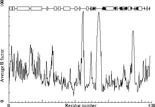
Chain without chain identifier
Hydrogen bond related checks
Error: HIS, ASN, GLN side chain flips
Listed here are Histidine, Asparagine or Glutamine residues for
which the orientation determined from hydrogen bonding analysis are
different from the assignment given in the input. Either they could
form energetically more favorable hydrogen bonds if the terminal
group was rotated by 180 degrees, or there is no assignment in the
input file (atom type 'A') but an assignment could be made. If a
residue is marked ``flexible'' the flipped conformation is only
slightly better than the non-flipped conformation.
 186 HIS  ( 188 )
 217 HIS  ( 219 )
 356 ASN  ( 361 )
In the table below all normal histidine residues are listed. The assignment based on the geometry of the residue is listed first, together with the RMS Z-score for the fit to the Engh and Huber parameters. For all residues where the H-bond assignment is different, the assignment is listed in the last columns, together with its RMS Z-score to the Engh and Huber parameters.
As always, the RMS Z-scores should be close to 1.0 if the residues were restrained to the Engh and Huber parameters during refinement.
Please note that because the differences between the geometries of the different types are small it is possible that the geometric assignment given here does not correspond to the type used in refinement. This is especially true if the RMS Z-scores are much higher than 1.0.
If the two assignments differ, or the ``geometry'' RMS Z-score is high, it is advisable to verify the hydrogen bond assignment, check the HIS type used during the refinement and possibly adjust it.
 137 HIS  ( 139 )     HIS-E   0.74 HIS-D   0.80
 171 HIS  ( 173 )     HIS-D   0.76 HIS-E   0.90
 182 HIS  ( 184 )     HIS-D   0.79 HIS-E   0.91
 186 HIS  ( 188 )     HIS-D   0.79
 199 HIS  ( 201 )     HIS-D   0.71 HIS-E   0.89
 217 HIS  ( 219 )     HIS-D   0.70 HIS-E   0.85
 258 HIS  ( 263 )     HIS-D   0.77 HIS-E   0.89
 280 HIS  ( 285 )     HIS-D   0.70
 306 HIS  ( 311 )     HIS-E   0.77
 381 HIS  ( 386 )     HIS-E   0.71
Hydrogen bond donors that are buried inside the protein normally use all of their hydrogens to form hydrogen bonds within the protein. If there are any non hydrogen bonded buried hydrogen bond donors in the structure they will be listed here. In very good structures the number of listed atoms will tend to zero.
   1 MET  (   1 )      N
  20 ASN  (  22 )      N
  21 PHE  (  23 )      N
  42 THR  (  44 )      OG1
 114 ASP  ( 116 )      N
 118 ASN  ( 120 )      N
 138 ILE  ( 140 )      N
 147 LEU  ( 149 )      N
 160 PHE  ( 162 )      N
 210 LYS  ( 212 )      N
 216 PHE  ( 218 )      N
 232 THR  ( 237 )      N
 233 ALA  ( 238 )      N
 265 GLU  ( 270 )      N
 274 ARG  ( 279 )      NH2
 275 GLY  ( 280 )      N
 276 LEU  ( 281 )      N
 281 GLN  ( 286 )      N
 319 ARG  ( 324 )      NE
 335 THR  ( 340 )      N
 347 THR  ( 352 )      N
 348 TYR  ( 353 )      N
 359 ASP  ( 364 )      N
 361 GLN  ( 366 )      N
 362 ALA  ( 367 )      N
 364 ARG  ( 369 )      NE
 364 ARG  ( 369 )      NH2
 372 LYS  ( 377 )      N
 374 ASP  ( 379 )      N
 375 LYS  ( 380 )      N
 376 LYS  ( 381 )      N
 413 GLU  ( 418 )      N
Side-chain hydrogen bond acceptors that are buried inside the protein normally form hydrogen bonds within the protein. If there are any not hydrogen bonded in the optimized hydrogen bond network they will be listed here.
  79 GLN  (  81 )      OE1
 286 GLU  ( 291 )      OE1
The second part of the table mostly gives an impression of how well the model conforms to common refinement constraint values. The first part of the table shows a number of constraint-independent quality indicators.
Structure Z-scores, positive is better than average:
  1st generation packing quality :  -0.633
  2nd generation packing quality :  -0.301
  Ramachandran plot appearance   :  -1.642
  chi-1/chi-2 rotamer normality  :  -2.083
  Backbone conformation          :  -0.169
  Bond lengths                   :   0.766
  Bond angles                    :   0.886
  Omega angle restraints         :   0.617 (tight)
  Side chain planarity           :   0.052 (tight)
  Improper dihedral distribution :   0.528
  B-factor distribution          :   1.515 (loose)
  Inside/Outside distribution    :   1.029