Improper dihedral RMS Z-score : 0.508
Note: Chain names are OK
All chain names assigned to polymer molecules are unique, and all
residue numbers are strictly increasing within each chain.
Note: Weights checked OK
All atomic occupancy factors ('weights') fall in the 0.0--1.0 range.
Geometric checks
Note: No missing atoms detected
All expected atoms are present.
Warning: C-terminal oxygen atoms missing
The C-atoms listed in the table below belong to a C-terminal residue
in a protein chain, but the C-terminal oxygen ("O2" or "OXT") that it
should be bound to was not found.
418 ILE ( 429 ) C
RMS Z-score for bond lengths: 0.763
RMS-deviation in bond distances: 0.017
Note: No bond length directionality
Comparison of bond distances with Engh and Huber [REF] standard
values for protein residues and Parkinson et al [REF] values for
DNA/RNA does not show significant systematic deviations.
Note: All bond angles OK
All bond angles are in agreement with standard bond angles using a
tolerance of 4 sigma (both standard values and sigma for protein
residues have been taken from Engh and Huber [REF], for DNA/RNA
from Parkinson et al. [REF]). Please note that only bond angles
within protein residues are taken into account: disulphide bridges
and peptide bonds are neglected.
Note: Normal bond angle variability
Bond angles were found to deviate normally from the mean standard
bond angles (normal values for protein residues were taken from
Engh and Huber [REF], for DNA/RNA from Parkinson et al [REF]). The
RMS Z-score given below is expected to be around 1.0 for a normally
restrained data set, and this is indeed observed for very high
resolution X-ray structures. More common values are around 1.55
RMS Z-score for bond angles: 0.901
RMS-deviation in bond angles: 1.806
Note: Side chain planarity OK
All of the side chains of residues that have a planar group are
planar within expected RMS deviations.
Note: Atoms connected to aromatic rings OK
All of the atoms that are connected to planar aromatic rings in side
chains of amino-acid residues are in the plane within expected RMS
deviations.
Warning: Unusual PRO puckering amplitudes
The proline residues listed in the table below have a puckering
amplitude that is outside of normal ranges. Puckering parameters
were calculated by the method of Cremer and Pople [REF]. Normal PRO
rings have a puckering amplitude Q between 0.20 and 0.45
Angstrom. If Q is lower than 0.20 Angstrom for a PRO residue, this
could indicate disorder between the two different normal ring forms
(with C-gamma below and above the ring, respectively). If Q is
higher than 0.45 Angstrom something could have gone wrong during the
refinement.
 103 PRO  ( 107 )     0.13 LOW
 227 PRO  ( 236 )     0.16 LOW
 256 PRO  ( 265 )     0.16 LOW
  11 PRO  (  11 )     99.6 envelop C-beta (108 degrees)
 191 PRO  ( 198 )    -21.2 half-chair C-alpha/N (-18 degrees)
 339 PRO  ( 348 )    102.6 envelop C-beta (108 degrees)
These scores give an impression of how ``normal'' the torsion angles in protein residues are. All torsion angles except omega are used for calculating a `normality' score. Average values and standard deviations were obtained from the residues in the WHAT IF database. These are used to calculate Z-scores. A residue with a Z-score of below -2.0 is poor, and a score of less than -3.0 is worrying. For such residues more than one torsion angle is in a highly unlikely position.
 339 PRO  ( 348 )   -2.7713
 228 THR  ( 237 )   -2.4351
 382 LEU  ( 393 )   -2.3378
 192 VAL  ( 199 )   -2.3084
 141 ASP  ( 147 )   -2.2900
 351 PHE  ( 362 )   -2.2111
 381 GLY  ( 392 )   -2.0921
 413 GLY  ( 424 )   -2.0664
Residues with ``forbidden'' phi-psi combinations are listed, as well as residues with unusual omega angles (deviating by more than 3 sigma from the normal value). Please note that it is normal if about 5 percent of the residues is listed here as having unusual phi-psi combinations.
 142 LEU  ( 148 )   omega poor
 152 SER  ( 158 )   Poor phi/psi
 188 PRO  ( 195 )   Poor PRO-phi, PRO omega poor
 189 PRO  ( 196 )   Poor PRO-phi
 191 PRO  ( 198 )   Poor PRO-phi
 203 LEU  ( 210 )   PRO omega poor
 204 PRO  ( 211 )   Poor PRO-phi
 247 LEU  ( 256 )   PRO omega poor
 248 PRO  ( 257 )   Poor PRO-phi
 266 GLY  ( 275 )   Poor phi/psi
 267 THR  ( 276 )   Poor phi/psi
 270 ARG  ( 279 )   Poor phi/psi
 271 GLY  ( 280 )   Poor phi/psi
 311 GLU  ( 320 )   Poor phi/psi
 339 PRO  ( 348 )   Poor PRO-phi
 340 GLY  ( 349 )   Poor phi/psi
 345 VAL  ( 356 )   Poor phi/psi
 352 GLU  ( 363 )   Poor phi/psi
 366 GLU  ( 377 )   Poor phi/psi
 369 GLU  ( 380 )   Poor phi/psi
 381 GLY  ( 392 )   omega poor
 414 GLY  ( 425 )   Poor phi/psi
Ramachandran Z-score : -1.588
Warning: Omega angles too tightly restrained
The omega angles for trans-peptide bonds in a structure are
expected to give a gaussian distribution with the average around
+178 degrees and a standard deviation around 5.5 degrees. These
expected values were obtained from very accurately determined
structures. Many protein structures are too tightly constrained.
This seems to be the case with the current structure, as the
observed standard deviation is below 4.0 degrees.
Standard deviation of omega values : 3.621
Note: chi-1/chi-2 angle correlation Z-score OK
The score expressing how well the chi-1/chi-2 angles of all residues
are corresponding to the populated areas in the database is
within expected ranges for well-refined structures.
chi-1/chi-2 correlation Z-score : -0.823
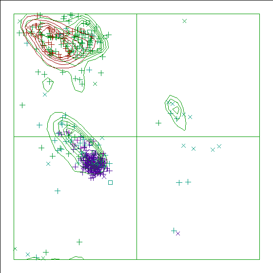
Note: Ramachandran plot
In this Ramachandran plot X-signs represent glycines, squares represent
prolines and small plus-signs represent the other residues. If too many
plus-signs fall outside the contoured areas then the molecule is poorly
refined (or worse).
In a colour picture, the residues that are part of a helix are shown in blue, strand residues in red. "Allowed" regions for helical residues are drawn in blue, for strand residues in red, and for all other residues in green.
Chain without chain identifier
Accessibility related checks
Note: Inside/Outside residue distribution normal
The distribution of residue types over the inside and the outside of the
protein is normal.
inside/outside RMS Z-score : 1.052
Note: Inside/Outside RMS Z-score plot
The Inside/Outside distribution normality RMS Z-score over a 15
residue window is plotted as function of the residue number. High
areas in the plot (above 1.5) indicate unusual inside/outside
patterns.
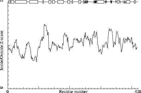
Chain without chain identifier
Secondary structure
Note: Secondary structure
This is the secondary structure according to DSSP. Only helix (H), strand
(S), turn (T) and coil (blank) are shown. [REF]
DBG> SSBOND cards to be written: 0
DBG> SSBOND cards to be written: 0
DBG> SSBOND cards to be written: 0
DBG> SSBOND cards to be written: 0
DBG> SSBOND cards to be written: 0
DBG> SSBOND cards to be written: 0
DBG> SSBOND cards to be written: 0
                     10        20
                      |         |
    1 -  22  MLSRQFVREHPETVRDAIERKG
    1 -  22     HHHHHH HHHHHHHHHTT
                   30        40        50        60
                    |         |         |         |
   23 -  69  VDLDEILDIDEEWRELKAEGDGLRQERNEVSSKIGELKQDGKDEEAQ
   23 -  69    HHHHHHHHHHHHHHHHHHHHHHHHHHHHHHHHHHHHHTT   TT
            70        80        90       100       110       120
             |         |         |         |         |         |
   70 - 124  RSQELKDELQDIEERADELESQLEEALLELPNIPHESVPTGEGEADNVERYREGF
   70 - 124   HHHHHHHHHHHHHHHHHHHHHHHHHHHT      TTT   TT333
                130       140       150       160       170       180
                  |         |         |         |         |         |
  125 - 181  DLPDEVVPHYDLGEDLDLLDFERGAKVSGGGYQFVKGEGARLEHALIQFMLDVHREQ
  125 - 181    TT    HHHHHHHTT   HHHHHHHT TT     HHHHHHHHHHHHHHHHHHHH
                   190       200       210
                     |         |         |
  182 - 219  EYVDVLPPIPVNSDSMEGTGQLPKFAEDAYRVGARQDD
  182 - 219          TT  HHHHHHHT TTTT333T
           220       230       240       250       260       270
             |         |         |         |         |         |
  220 - 279  SDDLWLLPTAEVPVTNMYRGEILLDDDLPVKHQAFSPNFRREAGEHGTETRGYVRVHQFH
  220 - 279         T THHHHHHTTTT    333  SSSSSSSSSS T      TT  T    TSSS
           280       290       300       310       320       330
             |         |         |         |         |         |
  280 - 339  KVELVNFVRPENSYDRLESLLDEAAEVLDRLELPYRVLDMCTGDMGFTQAKKYDIEVWAP
  280 - 339  SSSSSSS  333HHHHHHHHHHHHHHHHHHHT  SSSSS  TTTT TT TT SSSSS  T
           340
             |
  340 - 341  GD
  340 - 341  T
                   350       360       370       380       390       400
                     |         |         |         |         |         |
  342 - 401  WLEVSSVSNFEDFQARRAGLRYRPERHESADYLHTLNGSGLAVPRVLVAIMEYYQNDDGT
  342 - 401        SSS TTHHHHHHT SS  TTT   SS   SSS T  HHHHHHHHHHHT  TTT
                   410
                     |
  402 - 418  ITVPEPLRPYMGGQEVI
  402 - 418      TTT333TTT
The contact distances of all atom pairs have been checked. Two atoms are said to `bump' if they are closer than the sum of their Van der Waals radii minus 0.40 Angstrom. For hydrogen bonded pairs a tolerance of 0.55 Angstrom is used. The first number in the table tells you how much shorter that specific contact is than the acceptable limit. The second distance is the distance between the centers of the two atoms.
The last text-item on each line represents the status of the atom pair. The text `INTRA' means that the bump is between atoms that are explicitly listed in the PDB file. `INTER' means it is an inter-symmetry bump. If the final column contains the text 'HB', the bump criterium was relaxed because there could be a hydrogen bond. Similarly relaxed criteria are used for 1--3 and 1--4 interactions (listed as 'B2' and 'B3', respectively). If the last column is 'BF', the sum of the B-factors of the atoms is higher than 80, which makes the appearance of the bump somewhat less severe because the atoms probably aren't there anyway.
Bumps between atoms for which the sum of their occupancies is lower than one are not reported. In any case, each bump is listed in only one direction.
 365 PRO  ( 376 )      CD   --  370 SER  ( 381 )      O      0.892   1.908 INTRA
 190 ILE  ( 197 )      N    --  191 PRO  ( 198 )      CD     0.835   2.165 INTRA
 351 PHE  ( 362 )      CE2  --  378 ASN  ( 389 )      OD1    0.698   2.102 INTRA
 237 TYR  ( 246 )      CB   --  361 LEU  ( 372 )      CD2    0.610   2.590 INTRA
 223 LEU  ( 232 )      C    --  224 TRP  ( 233 )      CD1    0.499   2.701 INTRA
 327 THR  ( 336 )      CB   --  351 PHE  ( 362 )      CE1    0.489   2.711 INTRA
 237 TYR  ( 246 )      O    --  361 LEU  ( 372 )      CG     0.426   2.374 INTRA
 363 TYR  ( 374 )      CZ   --  372 ASP  ( 383 )      CB     0.415   2.785 INTRA
 351 PHE  ( 362 )      CZ   --  378 ASN  ( 389 )      OD1    0.409   2.391 INTRA
 189 PRO  ( 196 )      C    --  191 PRO  ( 198 )      CD     0.366   2.834 INTRA
  10 HIS  (  10 )      CB   --   13 THR  (  13 )      OG1    0.354   2.446 INTRA
  10 HIS  (  10 )      O    --   11 PRO  (  11 )      C      0.350   2.450 INTRA BF
 143 LEU  ( 149 )      CD2  --  145 PHE  ( 151 )      CE2    0.341   2.859 INTRA
 307 LEU  ( 316 )      O    --  311 GLU  ( 320 )      N      0.303   2.247 INTRA HB
 336 VAL  ( 345 )      CG2  --  345 VAL  ( 356 )      CG1    0.292   2.908 INTRA
 308 ASP  ( 317 )      O    --  311 GLU  ( 320 )      N      0.275   2.425 INTRA
 336 VAL  ( 345 )      CG2  --  345 VAL  ( 356 )      CG2    0.270   2.930 INTRA
 237 TYR  ( 246 )      C    --  361 LEU  ( 372 )      CD1    0.265   2.935 INTRA
 363 TYR  ( 374 )      CE2  --  372 ASP  ( 383 )      CB     0.257   2.943 INTRA
 414 GLY  ( 425 )      O    --  415 GLN  ( 426 )      C      0.254   2.546 INTRA
 140 LEU  ( 146 )      CB   --  142 LEU  ( 148 )      CG     0.246   2.954 INTRA
 327 THR  ( 336 )      O    --  351 PHE  ( 362 )      CD1    0.234   2.566 INTRA
 320 CYS  ( 329 )      O    --  321 THR  ( 330 )      C      0.229   2.571 INTRA
  10 HIS  (  10 )      N    --   11 PRO  (  11 )      CD     0.217   2.783 INTRA BF
 350 ASN  ( 361 )      ND2  --  352 GLU  ( 363 )      N      0.213   2.787 INTRA
And so on for a total of 182 lines
The packing environment of the residues is compared with the average packing environment for all residues of the same type in good PDB files. A low packing score can indicate one of several things: Poor packing, misthreading of the sequence through the density, crystal contacts, contacts with a co-factor, or the residue is part of the active site. It is not uncommon to see a few of these, but in any case this requires further inspection of the residue.
 216 ARG  ( 223 )    -7.91
 367 ARG  ( 378 )    -7.45
 270 ARG  ( 279 )    -7.26
 187 LEU  ( 194 )    -7.06
 274 ARG  ( 283 )    -6.87
 264 GLU  ( 273 )    -6.42
 217 GLN  ( 224 )    -5.85
 111 GLU  ( 115 )    -5.77
  61 GLN  (  63 )    -5.70
 119 ARG  ( 123 )    -5.68
 268 GLU  ( 277 )    -5.43
 358 ARG  ( 369 )    -5.33
 180 GLU  ( 186 )    -5.07
   9 GLU  (   9 )    -5.02
The table below lists the first and last residue in each stretch found, as well as the average residue score of the series.
216 ARG ( 223 ) --- 219 ASP ( 226 ) -5.73
Average for range 1 - 418 : -0.765
Note: Quality value plot
The quality value smoothed over a 10 residue window is plotted as
function of the residue number. Low areas in the plot (below
-2.0) indicate "unusual" packing.
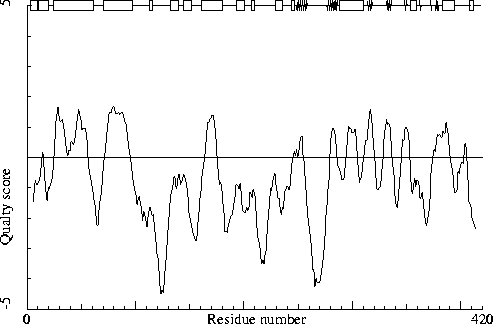
Chain without chain identifier
Warning: Low packing Z-score for some residues
The residues listed in the table below have an unusual packing
environment according to the 2nd generation quality check. The score
listed in the table is a packing normality Z-score: positive means
better than average, negative means worse than average. Only residues
scoring less than -2.50 are listed here. These are the "unusual"
residues in the structure, so it will be interesting to take a
special look at them.
 124 PHE  ( 128 )    -2.87
 372 ASP  ( 383 )    -2.78
 128 ASP  ( 134 )    -2.61
 236 MET  ( 245 )    -2.57
The table below lists the first and last residue in each stretch found, as well as the average residue Z-score of the series.
270 ARG ( 279 ) --- 275 VAL ( 284 ) -2.18
All contacts : Average = -0.058 Z-score = -0.23
BB-BB contacts : Average = 0.209 Z-score = 1.50
BB-SC contacts : Average = -0.362 Z-score = -1.90
SC-BB contacts : Average = 0.162 Z-score = 1.15
SC-SC contacts : Average = -0.255 Z-score = -1.09
Note: Second generation quality Z-score plot
The second generation quality Z-score smoothed over a 10 residue window
is plotted as function of the residue number. Low areas in the plot (below
-1.3) indicate "unusual" packing.
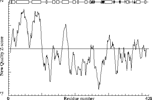
Chain without chain identifier
Warning: Backbone oxygen evaluation
The residues listed in the table below have an unusual backbone
oxygen position.
For each of the residues in the structure, a search was performed to find 5-residue stretches in the WHAT IF database with superposable C-alpha coordinates, and some constraints on the neighboring backbone oxygens.
In the following table the RMS distance between the backbone oxygen positions of these matching structures in the database and the position of the backbone oxygen atom in the current residue is given. If this number is larger than 1.5 a significant number of structures in the database show an alternative position for the backbone oxygen. If the number is larger than 2.0 most matching backbone fragments in the database have the peptide plane flipped. A manual check needs to be performed to assess whether the experimental data can support that alternative as well. The number in the last column is the number of database hits (maximum 80) used in the calculation. It is "normal" that some glycine residues show up in this list, but they are still worth checking!
154 GLY ( 160 ) 3.13 13
It is not necessarily an error if a few residues have rotamer values below 0.3, but careful inspection of all residues with these low values could be worth it.
 378 ASN  ( 389 )     0.38
 280 LYS  ( 289 )     0.40
For this check, backbone conformations are compared with database structures using C-alpha superpositions with some restraints on the backbone oxygen positions.
A residue mentioned in the table can be part of a strange loop, or there might be something wrong with it or its directly surrounding residues. There are a few of these in every protein, but in any case it is worth looking at!
 152 SER  ( 158 )    0
 191 PRO  ( 198 )    0
 203 LEU  ( 210 )    0
 267 THR  ( 276 )    0
 366 GLU  ( 377 )    0
 368 HIS  ( 379 )    0
 382 LEU  ( 393 )    0
 160 LYS  ( 166 )    1
 247 LEU  ( 256 )    1
 352 GLU  ( 363 )    1
 128 ASP  ( 134 )    2
 142 LEU  ( 148 )    2
 217 GLN  ( 224 )    2
 351 PHE  ( 362 )    2
Backbone conformation Z-score : 0.408
B-factor analysis
Note: Average B-factor OK
The average B-factor of buried atoms is within expected values for
a room-temperature X-ray study.
Average B-factor for buried atoms : 22.795
Note: Number of buried atoms with low B-factor is OK
For protein structures determined at room temperature, no more than
about 1 percent of the B factors of buried atoms is below 5.0.
Percentage of buried atoms with B less than 5 : 0.00
Error: The B-factors of bonded atoms show signs of over-refinement
For each of the bond types in a protein a distribution was derived
for the difference between the square roots of the B-factors of the
two atoms. All bonds in the current protein were scored against
these distributions. The number given below is the RMS Z-score over
the structure. For a structure with completely restrained B-factors
within residues, this value will be around 0.35, for extremely high
resolution structures refined with free isotropic B-factors this
number is expected to be near 1.0. Any value over 1.5 is sign of
severe over-refinement of B-factors.
RMS Z-score : 1.986 over 2911 bonds
Average difference in B over a bond : 2.39
RMS difference in B over a bond : 6.04
Note: B-factor plot
The average atomic B-factor per residue is plotted as function of
the residue number.
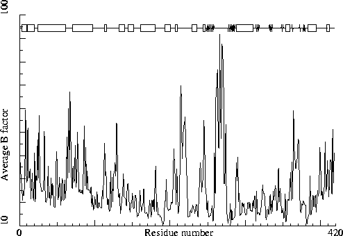
Chain without chain identifier
Hydrogen bond related checks
Error: HIS, ASN, GLN side chain flips
Listed here are Histidine, Asparagine or Glutamine residues for
which the orientation determined from hydrogen bonding analysis are
different from the assignment given in the input. Either they could
form energetically more favorable hydrogen bonds if the terminal
group was rotated by 180 degrees, or there is no assignment in the
input file (atom type 'A') but an assignment could be made. If a
residue is marked ``flexible'' the flipped conformation is only
slightly better than the non-flipped conformation.
350 ASN ( 361 )
In the table below all normal histidine residues are listed. The assignment based on the geometry of the residue is listed first, together with the RMS Z-score for the fit to the Engh and Huber parameters. For all residues where the H-bond assignment is different, the assignment is listed in the last columns, together with its RMS Z-score to the Engh and Huber parameters.
As always, the RMS Z-scores should be close to 1.0 if the residues were restrained to the Engh and Huber parameters during refinement.
Please note that because the differences between the geometries of the different types are small it is possible that the geometric assignment given here does not correspond to the type used in refinement. This is especially true if the RMS Z-scores are much higher than 1.0.
If the two assignments differ, or the ``geometry'' RMS Z-score is high, it is advisable to verify the hydrogen bond assignment, check the HIS type used during the refinement and possibly adjust it.
  10 HIS  (  10 )     HIS-E   0.88
 104 HIS  ( 108 )     HIS-E   0.77
 133 HIS  ( 139 )     HIS-E   0.64 HIS-D   0.81
 168 HIS  ( 174 )     HIS-E   0.66 HIS-D   0.95
 178 HIS  ( 184 )     HIS-D   0.80 HIS-E   0.89
 251 HIS  ( 260 )     HIS-E   0.80 HIS-H   1.03
 265 HIS  ( 274 )     HIS-D   0.70
 276 HIS  ( 285 )     HIS-D   0.79
 279 HIS  ( 288 )     HIS-D   0.76
 368 HIS  ( 379 )     HIS-D   0.81 HIS-E   0.90
 375 HIS  ( 386 )     HIS-E   0.77
Hydrogen bond donors that are buried inside the protein normally use all of their hydrogens to form hydrogen bonds within the protein. If there are any non hydrogen bonded buried hydrogen bond donors in the structure they will be listed here. In very good structures the number of listed atoms will tend to zero.
   1 MET  (   1 )      N
  13 THR  (  13 )      N
  20 ARG  (  20 )      NE
 111 GLU  ( 115 )      N
 116 ASN  ( 120 )      N
 134 TYR  ( 140 )      N
 143 LEU  ( 149 )      N
 156 TYR  ( 162 )      N
 205 LYS  ( 212 )      N
 211 TYR  ( 218 )      N
 228 THR  ( 237 )      N
 245 ASP  ( 254 )      N
 252 GLN  ( 261 )      NE2
 277 GLN  ( 286 )      N
 279 HIS  ( 288 )      N
 279 HIS  ( 288 )      ND1
 285 ASN  ( 294 )      ND2
 294 ASP  ( 303 )      N
 315 ARG  ( 324 )      NE
 331 LYS  ( 340 )      N
 353 ASP  ( 364 )      N
 355 GLN  ( 366 )      N
 358 ARG  ( 369 )      NE
 358 ARG  ( 369 )      NH2
 366 GLU  ( 377 )      N
 368 HIS  ( 379 )      N
 369 GLU  ( 380 )      N
 370 SER  ( 381 )      N
 386 ARG  ( 397 )      N
 396 GLN  ( 407 )      N
 402 ILE  ( 413 )      N
Side-chain hydrogen bond acceptors that are buried inside the protein normally form hydrogen bonds within the protein. If there are any not hydrogen bonded in the optimized hydrogen bond network they will be listed here.
 167 GLU  ( 173 )      OE2
 252 GLN  ( 261 )      OE1
 378 ASN  ( 389 )      OD1
The second part of the table mostly gives an impression of how well the model conforms to common refinement constraint values. The first part of the table shows a number of constraint-independent quality indicators.
Structure Z-scores, positive is better than average:
  1st generation packing quality :  -0.662
  2nd generation packing quality :  -0.229
  Ramachandran plot appearance   :  -1.588
  chi-1/chi-2 rotamer normality  :  -0.823
  Backbone conformation          :   0.408
  Bond lengths                   :   0.763
  Bond angles                    :   0.901
  Omega angle restraints         :   0.658 (tight)
  Side chain planarity           :   0.056 (tight)
  Improper dihedral distribution :   0.508
  B-factor distribution          :   1.986 (loose)
  Inside/Outside distribution    :   1.052