Improper dihedrals are a measure of the chirality/planarity of the structure at a specific atom. Values around -35 or +35 are expected for chiral atoms, and values around 0 for planar atoms. Planar side chains are left out of the calculations, these are better handled by the planarity checks.
Three numbers are given for each atom in the table. The first is the Z-score for the improper dihedral. The second number is the measured improper dihedral. The third number is the expected value for this atom type. A final column contains an extra warning if the chirality for an atom is opposite to the expected value.
 405 ILE  ( 423 )      C        5.3     10.3     -0.2
 406 PRO  ( 424 )      N        7.2     27.8     -1.5
Improper dihedral RMS Z-score : 0.562
Note: Chain names are OK
All chain names assigned to polymer molecules are unique, and all
residue numbers are strictly increasing within each chain.
Note: Weights checked OK
All atomic occupancy factors ('weights') fall in the 0.0--1.0 range.
Geometric checks
Note: No missing atoms detected
All expected atoms are present.
Warning: C-terminal oxygen atoms missing
The C-atoms listed in the table below belong to a C-terminal residue
in a protein chain, but the C-terminal oxygen ("O2" or "OXT") that it
should be bound to was not found.
407 GLY ( 425 ) C
Atom names starting with "<" belong to the previous residue in the chain. If the second atom name is "--SS", the disulphide bridge has a deviating length.
406 PRO ( 424 ) CD N 1.336 -9.8
RMS Z-score for bond lengths: 0.776
RMS-deviation in bond distances: 0.017
Note: No bond length directionality
Comparison of bond distances with Engh and Huber [REF] standard
values for protein residues and Parkinson et al [REF] values for
DNA/RNA does not show significant systematic deviations.
Warning: Unusual bond angles
The bond angles listed in the table below were found to deviate
more than 4 sigma from standard bond angles (both standard values
and sigma for protein residues have been taken from Engh and Huber
[REF], for DNA/RNA from Parkinson et al [REF]). In the table below
for each strange angle the bond angle and the number of standard
deviations it differs from the standard values is given. Please
note that disulphide bridges are neglected. Atoms starting with "<"
belong to the previous residue in the sequence.
 264 TRP  ( 279 )      C    CA   CB  119.417   4.9
 406 PRO  ( 424 )      CG   CD   N    95.833  -4.9
RMS Z-score for bond angles: 0.894
RMS-deviation in bond angles: 1.783
Note: Side chain planarity OK
All of the side chains of residues that have a planar group are
planar within expected RMS deviations.
Note: Atoms connected to aromatic rings OK
All of the atoms that are connected to planar aromatic rings in side
chains of amino-acid residues are in the plane within expected RMS
deviations.
Warning: Unusual PRO puckering amplitudes
The proline residues listed in the table below have a puckering
amplitude that is outside of normal ranges. Puckering parameters
were calculated by the method of Cremer and Pople [REF]. Normal PRO
rings have a puckering amplitude Q between 0.20 and 0.45
Angstrom. If Q is lower than 0.20 Angstrom for a PRO residue, this
could indicate disorder between the two different normal ring forms
(with C-gamma below and above the ring, respectively). If Q is
higher than 0.45 Angstrom something could have gone wrong during the
refinement.
 144 PRO  ( 151 )     0.14 LOW
 184 PRO  ( 192 )     0.11 LOW
 188 PRO  ( 196 )     0.12 LOW
 406 PRO  ( 424 )     0.50 HIGH
 126 PRO  ( 128 )    -24.0 half-chair C-alpha/N (-18 degrees)
 333 PRO  ( 348 )    103.3 envelop C-beta (108 degrees)
 406 PRO  ( 424 )   -153.0 half-chair N/C-delta (-162 degrees)
These scores give an impression of how ``normal'' the torsion angles in protein residues are. All torsion angles except omega are used for calculating a `normality' score. Average values and standard deviations were obtained from the residues in the WHAT IF database. These are used to calculate Z-scores. A residue with a Z-score of below -2.0 is poor, and a score of less than -3.0 is worrying. For such residues more than one torsion angle is in a highly unlikely position.
 406 PRO  ( 424 )   -3.0369
 332 PHE  ( 347 )   -2.9490
 348 THR  ( 363 )   -2.8116
 333 PRO  ( 348 )   -2.8071
 126 PRO  ( 128 )   -2.5930
 129 LEU  ( 134 )   -2.5899
 224 THR  ( 237 )   -2.3671
 185 LEU  ( 193 )   -2.2556
 405 ILE  ( 423 )   -2.2318
 189 VAL  ( 197 )   -2.2133
 140 ASP  ( 147 )   -2.1534
 266 VAL  ( 281 )   -2.0438
 148 VAL  ( 155 )   -2.0237
 112 SER  ( 114 )   -2.0125
 355 LEU  ( 370 )   -2.0025
Residues with ``forbidden'' phi-psi combinations are listed, as well as residues with unusual omega angles (deviating by more than 3 sigma from the normal value). Please note that it is normal if about 5 percent of the residues is listed here as having unusual phi-psi combinations.
  10 ASP  (  10 )   Poor phi/psi
  20 ALA  (  22 )   Poor phi/psi
  63 LYS  (  65 )   Poor phi/psi
 112 SER  ( 114 )   Poor phi/psi, omega poor
 125 LYS  ( 127 )   Poor phi/psi
 126 PRO  ( 128 )   Poor PRO-phi
 151 CYS  ( 158 )   Poor phi/psi
 154 ARG  ( 161 )   Poor phi/psi
 160 ASN  ( 167 )   Poor phi/psi
 200 ALA  ( 208 )   Poor phi/psi
 202 LEU  ( 210 )   omega poor
 218 GLU  ( 226 )   Poor phi/psi
 241 LEU  ( 256 )   PRO omega poor
 242 PRO  ( 257 )   Poor PRO-phi
 260 GLY  ( 275 )   Poor phi/psi
 261 LYS  ( 276 )   Poor phi/psi
 264 TRP  ( 279 )   Poor phi/psi
 265 GLY  ( 280 )   Poor phi/psi
 305 LYS  ( 320 )   Poor phi/psi
 333 PRO  ( 348 )   Poor PRO-phi
 334 TYR  ( 349 )   Poor phi/psi
 341 LEU  ( 356 )   Poor phi/psi
 348 THR  ( 363 )   Poor phi/psi
 356 GLU  ( 371 )   Poor phi/psi
 375 LEU  ( 392 )   omega poor
 406 PRO  ( 424 )   Poor phi/psi
Ramachandran Z-score : -1.756
Warning: Omega angles too tightly restrained
The omega angles for trans-peptide bonds in a structure are
expected to give a gaussian distribution with the average around
+178 degrees and a standard deviation around 5.5 degrees. These
expected values were obtained from very accurately determined
structures. Many protein structures are too tightly constrained.
This seems to be the case with the current structure, as the
observed standard deviation is below 4.0 degrees.
Standard deviation of omega values : 3.491
Note: chi-1/chi-2 angle correlation Z-score OK
The score expressing how well the chi-1/chi-2 angles of all residues
are corresponding to the populated areas in the database is
within expected ranges for well-refined structures.
chi-1/chi-2 correlation Z-score : -0.309
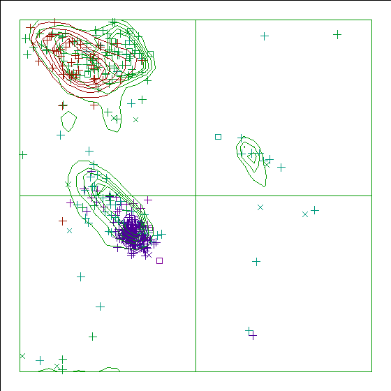
Note: Ramachandran plot
In this Ramachandran plot X-signs represent glycines, squares represent
prolines and small plus-signs represent the other residues. If too many
plus-signs fall outside the contoured areas then the molecule is poorly
refined (or worse).
In a colour picture, the residues that are part of a helix are shown in blue, strand residues in red. "Allowed" regions for helical residues are drawn in blue, for strand residues in red, and for all other residues in green.
Chain without chain identifier
Accessibility related checks
Note: Inside/Outside residue distribution normal
The distribution of residue types over the inside and the outside of the
protein is normal.
inside/outside RMS Z-score : 1.126
Note: Inside/Outside RMS Z-score plot
The Inside/Outside distribution normality RMS Z-score over a 15
residue window is plotted as function of the residue number. High
areas in the plot (above 1.5) indicate unusual inside/outside
patterns.
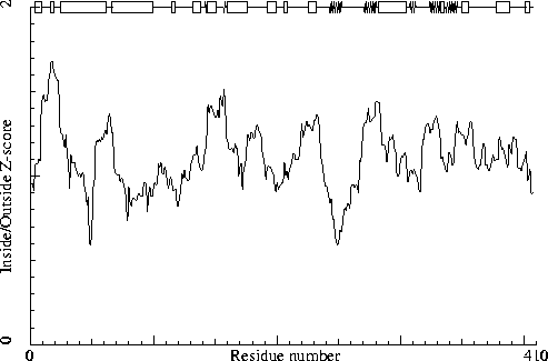
Chain without chain identifier
Secondary structure
Note: Secondary structure
This is the secondary structure according to DSSP. Only helix (H), strand
(S), turn (T) and coil (blank) are shown. [REF]
DBG> SSBOND cards to be written: 0
DBG> SSBOND cards to be written: 0
A molecule was found with too few intact residues to determine
the secondary structure
DBG> SSBOND cards to be written: 0
DBG> SSBOND cards to be written: 0
DBG> SSBOND cards to be written: 0
DBG> SSBOND cards to be written: 0
DBG> SSBOND cards to be written: 0
DBG> SSBOND cards to be written: 0
                     10
                      |
    1 -  15  MLDINQFIEDKGGNP
    1 -  15     HHHHHH TTT
                20        30        40        50        60        70
                 |         |         |         |         |         |
   16 -  75  KARNASVEIVDEIISDYKDWVKTRFELDELNKKFNKLQKDIGLKFKNKEDASGLLAEKEK
   16 -  75   333T    HHHHHHHHHHHHHHHHHHHHHHHHHHHHHHHHHHHHHTT   HHHHHHHHH
                80        90       100       110       120
                 |         |         |         |         |
   76 - 127  LTQQKKELTEKEQQEDKDLKKKVFQVGNIVHPSVVVSNDEENNELVRTWKPE
   76 - 127  HHHHHHHHHHHHHHHHHHHHHHHHT      TTT   TT333       T
             130
               |
  128 - 130  DLE
  128 - 130  ???
                    140       150       160       170
                      |         |         |         |
  131 - 176  SHHEILLRLDGYDPDRGVKICGHRGYFFRNYGVFLNQALINYGLQF
  131 - 176   HHHHHHHTT SSHHHHHHHT TT  SS HHHHHHHHHHHHHHHH
              180       190       200       210       220
                |         |         |         |         |
  177 - 220  LAAKGYIPLQAPVMMNKELMSKTAQLSEFDEELYKVIDGEDEKY
  177 - 220    TT        T   HHHHHHHT TTTT333T       TT
                    230
                      |
  221 - 238  LIATSEQPISAYHSGEWF
  221 - 238    T THHHHHHTTTT
            240       250       260       270       280       290
              |         |         |         |         |         |
  239 - 298  EQLPIHYVGYSSCFRREAGSHGKDAWGVFRVHAFEKIEQFVITEPEKSWEEFEKMISYSE
  239 - 298      SSSSSSSSSS T      TT  T    TSSSSSSSSSS  333HHHHHHHHHHHHH
            300       310       320       330       340       350
              |         |         |         |         |         |
  299 - 358  EFYKSLKLPYRIVGIVSGELNNAAAKKYDLEAWFPYQKEYKELVSCSNCTDYQSRNLEIR
  299 - 358  HHHHHHT  SSSSS  TTTT TT TSSSSSSSSS333TSSSSSSSSSS TTHHHHHHT
            360
              |
  359 - 362  CGIK
  359 - 362
                  370       380       390
                    |         |         |
  363 - 396  REKKYVHCLNSTLAATQRALCCILENYQTEDGLV
  363 - 396              T  HHHHHHHHHHHT  TTT
              400
                |
  397 - 407  VPEVLRKYIPG
  397 - 407    TTT333TT
The contact distances of all atom pairs have been checked. Two atoms are said to `bump' if they are closer than the sum of their Van der Waals radii minus 0.40 Angstrom. For hydrogen bonded pairs a tolerance of 0.55 Angstrom is used. The first number in the table tells you how much shorter that specific contact is than the acceptable limit. The second distance is the distance between the centers of the two atoms.
The last text-item on each line represents the status of the atom pair. The text `INTRA' means that the bump is between atoms that are explicitly listed in the PDB file. `INTER' means it is an inter-symmetry bump. If the final column contains the text 'HB', the bump criterium was relaxed because there could be a hydrogen bond. Similarly relaxed criteria are used for 1--3 and 1--4 interactions (listed as 'B2' and 'B3', respectively). If the last column is 'BF', the sum of the B-factors of the atoms is higher than 80, which makes the appearance of the bump somewhat less severe because the atoms probably aren't there anyway.
Bumps between atoms for which the sum of their occupancies is lower than one are not reported. In any case, each bump is listed in only one direction.
  11 LYS  (  11 )      O    --   15 PRO  (  15 )      CD     1.289   1.511 INTRA
 233 HIS  ( 246 )      ND1  --  238 PHE  ( 251 )      CZ     0.792   2.308 INTRA
  11 LYS  (  11 )      C    --   15 PRO  (  15 )      CD     0.632   2.568 INTRA
 225 SER  ( 238 )      OG   --  276 GLU  ( 291 )      CB     0.605   2.195 INTRA
  11 LYS  (  11 )      O    --   15 PRO  (  15 )      CG     0.597   2.203 INTRA
  12 GLY  (  12 )      C    --   15 PRO  (  15 )      CD     0.593   2.607 INTRA
 199 THR  ( 207 )      O    --  200 ALA  ( 208 )      CB     0.558   2.242 INTRA
 248 TYR  ( 263 )      OH   --  273 GLU  ( 288 )      CB     0.496   2.304 INTRA
 117 ASN  ( 119 )      O    --  314 VAL  ( 329 )      CG1    0.472   2.328 INTRA
 341 LEU  ( 356 )      CD1  --  384 CYS  ( 401 )      SG     0.460   2.940 INTRA
  12 GLY  (  12 )      C    --   15 PRO  (  15 )      CG     0.446   2.754 INTRA
  12 GLY  (  12 )      CA   --   15 PRO  (  15 )      CG     0.444   2.756 INTRA
 248 TYR  ( 263 )      CZ   --  273 GLU  ( 288 )      CB     0.429   2.771 INTRA
  12 GLY  (  12 )      O    --   15 PRO  (  15 )      CG     0.420   2.380 INTRA
 290 PHE  ( 305 )      O    --  294 ILE  ( 309 )      CG2    0.392   2.408 INTRA
 203 SER  ( 211 )      O    --  206 ASP  ( 214 )      OD1    0.387   2.013 INTRA
 344 CYS  ( 359 )      SG   --  373 SER  ( 390 )      CB     0.359   3.041 INTRA
 198 LYS  ( 206 )      CB   --  355 LEU  ( 370 )      CD2    0.356   2.844 INTRA
 290 PHE  ( 305 )      CZ   --  326 TYR  ( 341 )      CD2    0.355   2.845 INTRA
 293 MET  ( 308 )      CE   --  371 LEU  ( 388 )      CB     0.354   2.846 INTRA
 199 THR  ( 207 )      CG2  --  230 SER  ( 243 )      OG     0.349   2.451 INTRA
 266 VAL  ( 281 )      CG2  --  339 LYS  ( 354 )      CG     0.347   2.853 INTRA
 172 TYR  ( 179 )      CE1  --  400 VAL  ( 418 )      CG2    0.345   2.855 INTRA
 142 TYR  ( 149 )      CE1  --  158 PHE  ( 165 )      CZ     0.335   2.865 INTRA
 112 SER  ( 114 )      CB   --  117 ASN  ( 119 )      CB     0.333   2.867 INTRA
And so on for a total of 226 lines
The packing environment of the residues is compared with the average packing environment for all residues of the same type in good PDB files. A low packing score can indicate one of several things: Poor packing, misthreading of the sequence through the density, crystal contacts, contacts with a co-factor, or the residue is part of the active site. It is not uncommon to see a few of these, but in any case this requires further inspection of the residue.
 186 GLN  ( 194 )    -7.38
 129 LEU  ( 134 )    -6.50
 153 HIS  ( 160 )    -6.45
 159 ARG  ( 166 )    -6.32
 240 GLN  ( 255 )    -5.95
 268 ARG  ( 283 )    -5.92
 213 ILE  ( 221 )    -5.88
 335 GLN  ( 350 )    -5.49
  19 ASN  (  21 )    -5.27
 113 ASN  ( 115 )    -5.25
 392 GLU  ( 409 )    -5.12
The table below lists the first and last residue in each stretch found, as well as the average residue score of the series.
334 TYR ( 349 ) --- 336 LYS ( 351 ) -4.89
Average for range 1 - 407 : -0.738
Note: Quality value plot
The quality value smoothed over a 10 residue window is plotted as
function of the residue number. Low areas in the plot (below
-2.0) indicate "unusual" packing.
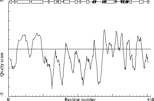
Chain without chain identifier
Warning: Low packing Z-score for some residues
The residues listed in the table below have an unusual packing
environment according to the 2nd generation quality check. The score
listed in the table is a packing normality Z-score: positive means
better than average, negative means worse than average. Only residues
scoring less than -2.50 are listed here. These are the "unusual"
residues in the structure, so it will be interesting to take a
special look at them.
 213 ILE  ( 221 )    -3.61
 355 LEU  ( 370 )    -2.78
 204 GLU  ( 212 )    -2.63
 129 LEU  ( 134 )    -2.57
 216 GLU  ( 224 )    -2.55
 265 GLY  ( 280 )    -2.55
 153 HIS  ( 160 )    -2.51
All contacts : Average = -0.175 Z-score = -1.01
BB-BB contacts : Average = 0.144 Z-score = 1.04
BB-SC contacts : Average = -0.367 Z-score = -1.93
SC-BB contacts : Average = 0.081 Z-score = 0.66
SC-SC contacts : Average = -0.349 Z-score = -1.60
Note: Second generation quality Z-score plot
The second generation quality Z-score smoothed over a 10 residue window
is plotted as function of the residue number. Low areas in the plot (below
-1.3) indicate "unusual" packing.
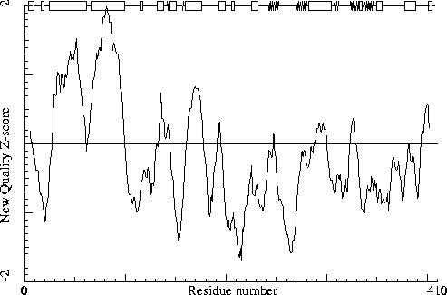
Chain without chain identifier
Warning: Backbone oxygen evaluation
The residues listed in the table below have an unusual backbone
oxygen position.
For each of the residues in the structure, a search was performed to find 5-residue stretches in the WHAT IF database with superposable C-alpha coordinates, and some constraints on the neighboring backbone oxygens.
In the following table the RMS distance between the backbone oxygen positions of these matching structures in the database and the position of the backbone oxygen atom in the current residue is given. If this number is larger than 1.5 a significant number of structures in the database show an alternative position for the backbone oxygen. If the number is larger than 2.0 most matching backbone fragments in the database have the peptide plane flipped. A manual check needs to be performed to assess whether the experimental data can support that alternative as well. The number in the last column is the number of database hits (maximum 80) used in the calculation. It is "normal" that some glycine residues show up in this list, but they are still worth checking!
215 GLY ( 223 ) 1.61 13
It is not necessarily an error if a few residues have rotamer values below 0.3, but careful inspection of all residues with these low values could be worth it.
 399 GLU  ( 417 )     0.33
 374 THR  ( 391 )     0.36
 274 LYS  ( 289 )     0.36
  30 SER  (  32 )     0.39
For this check, backbone conformations are compared with database structures using C-alpha superpositions with some restraints on the backbone oxygen positions.
A residue mentioned in the table can be part of a strange loop, or there might be something wrong with it or its directly surrounding residues. There are a few of these in every protein, but in any case it is worth looking at!
 125 LYS  ( 127 )    0
 151 CYS  ( 158 )    0
 161 TYR  ( 168 )    0
 200 ALA  ( 208 )    0
 202 LEU  ( 210 )    0
 261 LYS  ( 276 )    0
 348 THR  ( 363 )    0
 375 LEU  ( 392 )    0
 376 ALA  ( 393 )    0
 153 HIS  ( 160 )    1
 154 ARG  ( 161 )    1
 218 GLU  ( 226 )    1
 241 LEU  ( 256 )    1
  20 ALA  (  22 )    2
 111 VAL  ( 113 )    2
 160 ASN  ( 167 )    2
 216 GLU  ( 224 )    2
 347 CYS  ( 362 )    2
Backbone conformation Z-score : 0.110
B-factor analysis
Note: Average B-factor OK
The average B-factor of buried atoms is within expected values for
a room-temperature X-ray study.
Average B-factor for buried atoms : 22.397
Note: Number of buried atoms with low B-factor is OK
For protein structures determined at room temperature, no more than
about 1 percent of the B factors of buried atoms is below 5.0.
Percentage of buried atoms with B less than 5 : 0.00
Error: The B-factors of bonded atoms show signs of over-refinement
For each of the bond types in a protein a distribution was derived
for the difference between the square roots of the B-factors of the
two atoms. All bonds in the current protein were scored against
these distributions. The number given below is the RMS Z-score over
the structure. For a structure with completely restrained B-factors
within residues, this value will be around 0.35, for extremely high
resolution structures refined with free isotropic B-factors this
number is expected to be near 1.0. Any value over 1.5 is sign of
severe over-refinement of B-factors.
RMS Z-score : 2.051 over 2894 bonds
Average difference in B over a bond : 2.36
RMS difference in B over a bond : 6.03
Note: B-factor plot
The average atomic B-factor per residue is plotted as function of
the residue number.
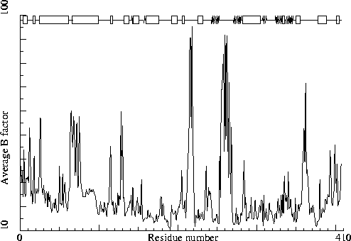
Chain without chain identifier
Hydrogen bond related checks
Error: HIS, ASN, GLN side chain flips
Listed here are Histidine, Asparagine or Glutamine residues for
which the orientation determined from hydrogen bonding analysis are
different from the assignment given in the input. Either they could
form energetically more favorable hydrogen bonds if the terminal
group was rotated by 180 degrees, or there is no assignment in the
input file (atom type 'A') but an assignment could be made. If a
residue is marked ``flexible'' the flipped conformation is only
slightly better than the non-flipped conformation.
  62 ASN  (  64 )
 106 HIS  ( 108 )
 259 HIS  ( 274 )
 346 ASN  ( 361 )
In the table below all normal histidine residues are listed. The assignment based on the geometry of the residue is listed first, together with the RMS Z-score for the fit to the Engh and Huber parameters. For all residues where the H-bond assignment is different, the assignment is listed in the last columns, together with its RMS Z-score to the Engh and Huber parameters.
As always, the RMS Z-scores should be close to 1.0 if the residues were restrained to the Engh and Huber parameters during refinement.
Please note that because the differences between the geometries of the different types are small it is possible that the geometric assignment given here does not correspond to the type used in refinement. This is especially true if the RMS Z-scores are much higher than 1.0.
If the two assignments differ, or the ``geometry'' RMS Z-score is high, it is advisable to verify the hydrogen bond assignment, check the HIS type used during the refinement and possibly adjust it.
 106 HIS  ( 108 )     HIS-H   0.68 HIS-E   0.76
 132 HIS  ( 139 )     HIS-E   0.80 HIS-D   0.82
 133 HIS  ( 140 )     HIS-D   0.79 HIS-E   0.89
 153 HIS  ( 160 )     HIS-E   0.86
 233 HIS  ( 246 )     HIS-E   0.85
 244 HIS  ( 259 )     HIS-E   0.77
 259 HIS  ( 274 )     HIS-D   0.78 HIS-E   0.83
 270 HIS  ( 285 )     HIS-D   0.83
 369 HIS  ( 386 )     HIS-E   0.75
Hydrogen bond donors that are buried inside the protein normally use all of their hydrogens to form hydrogen bonds within the protein. If there are any non hydrogen bonded buried hydrogen bond donors in the structure they will be listed here. In very good structures the number of listed atoms will tend to zero.
   1 MET  (   1 )      N
   2 LEU  (   2 )      N
 118 ASN  ( 120 )      N
 133 HIS  ( 140 )      N
 161 TYR  ( 168 )      N
 179 ALA  ( 187 )      N
 180 LYS  ( 188 )      N
 182 TYR  ( 190 )      N
 190 MET  ( 198 )      N
 204 GLU  ( 212 )      N
 210 TYR  ( 218 )      N
 218 GLU  ( 226 )      N
 223 ALA  ( 236 )      N
 224 THR  ( 237 )      N
 225 SER  ( 238 )      N
 233 HIS  ( 246 )      NE2
 234 SER  ( 247 )      OG
 265 GLY  ( 280 )      N
 266 VAL  ( 281 )      N
 277 GLN  ( 292 )      NE2
 282 GLU  ( 297 )      N
 301 TYR  ( 316 )      OH
 309 ARG  ( 324 )      NE
 319 ASN  ( 334 )      N
 337 GLU  ( 352 )      N
 338 TYR  ( 353 )      N
 340 GLU  ( 355 )      N
 343 SER  ( 358 )      N
 348 THR  ( 363 )      OG1
 349 ASP  ( 364 )      N
 351 GLN  ( 366 )      N
 352 SER  ( 367 )      OG
 358 ARG  ( 373 )      NE
 364 GLU  ( 381 )      N
 380 ARG  ( 397 )      N
 390 GLN  ( 407 )      N
 400 VAL  ( 418 )      N
 402 ARG  ( 420 )      N
Side-chain hydrogen bond acceptors that are buried inside the protein normally form hydrogen bonds within the protein. If there are any not hydrogen bonded in the optimized hydrogen bond network they will be listed here.
  14 ASN  (  14 )      OD1
  23 GLU  (  25 )      OE2
  87 GLU  (  89 )      OE1
 244 HIS  ( 259 )      ND1
 276 GLU  ( 291 )      OE1
 351 GLN  ( 366 )      OE1
The second part of the table mostly gives an impression of how well the model conforms to common refinement constraint values. The first part of the table shows a number of constraint-independent quality indicators.
Structure Z-scores, positive is better than average:
  1st generation packing quality :  -0.595
  2nd generation packing quality :  -1.006
  Ramachandran plot appearance   :  -1.756
  chi-1/chi-2 rotamer normality  :  -0.309
  Backbone conformation          :   0.110
  Bond lengths                   :   0.776
  Bond angles                    :   0.894
  Omega angle restraints         :   0.635 (tight)
  Side chain planarity           :   0.074 (tight)
  Improper dihedral distribution :   0.562
  B-factor distribution          :   2.051 (loose)
  Inside/Outside distribution    :   1.126