The short videos presented in this page are not a replacement of the seminar. They merely explain once more a few of the main points made in the validation seminar and practical. |
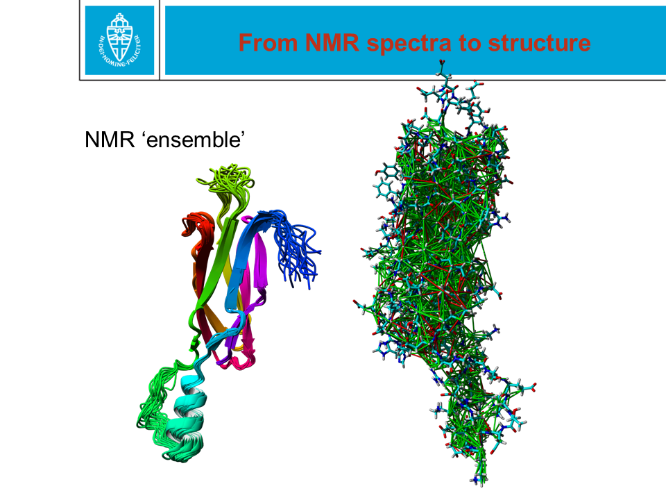
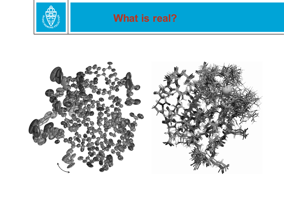
Figure 28. When a protein structure is being solved by X-ray or NMR, coordinates are obtained for the (most) atoms. These two methods are technologically and scientifically very different. In the PDB file, coordinates are given with an error margin of only 0.0005 Ångström. That precision is NOT realistic. |
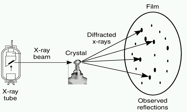
Figure 29. When you shoot X-rays at a protein crystal, you can obtain a diffraction pattern from which the coordinates of the atoms can be calculated. This easier said than done, though. |
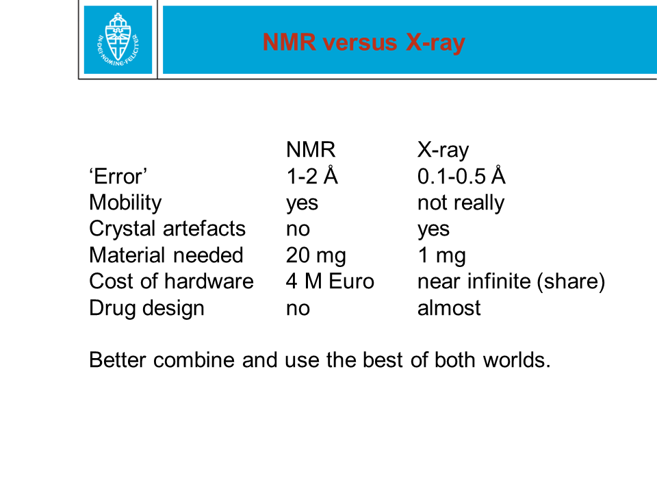
Figure 31. NMR and X-ray both have their own advantages and disadvantages. |
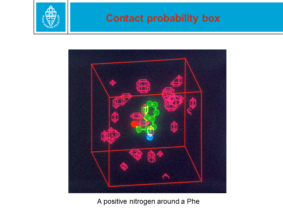
Figure 33. Many validation techniques rely on the use of Force Fields. Structure packing normality analysis is a nice example that we will come back to at a later stage. |
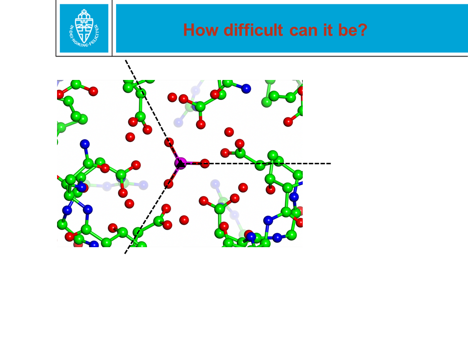
Figure 34. Many of the errors we showed are really big errors, and everybody who has worked for a few weeks with protein structures will spot them immediately. Other errors, though, can be hard to spot. This example is about a phosphate ion that is sitting at a special position. Two of the three crystallographers involved are among the best in the world. And even when we told them that there was a problem with this phosphate, they did not spot it. |
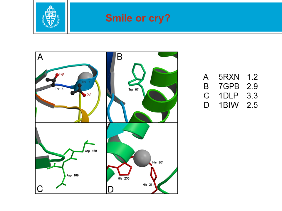
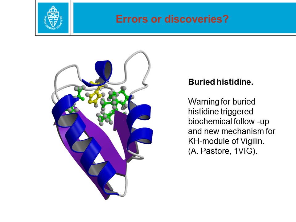
Figure 35. Often, the software flags things as abnormal. Abnormal is not wrong! In some cases, a warning for something abnormal can lead to nice science to answer relevant questions. |